💪🧬🥇NOW 7 INTERNATIONAL PUBLICATIONS (and more to come) TO ATTEST THE ROBUSTNESS AND EFFICIENCY OF EX VIVO CLINICAL APPROACH – CLINIC’N’CELL

https://pubmed.ncbi.nlm.nih.gov/37111121/

Nutrients 2023 Apr 14;15(8):1903. doi: 10.3390/nu15081903.

Circulating Human Metabolites Resulting from TOTUM-070 Absorption (a Plant-Based, Polyphenol-Rich Ingredient) Improve Lipid Metabolism in Human Hepatocytes: Lessons from an Original Ex Vivo Clinical Trial

Fabien Wauquier 1Line Boutin-Wittrant 1Stéphanie Krisa 2Josep Valls 2 3Cedric Langhi 4Yolanda F Otero 4Pascal Sirvent 4Sébastien Peltier 5Maxime Bargetto 5Murielle Cazaubiel 5Véronique Sapone 5Annie Bouchard-Mercier 6Véronique Roux 7Nicolas Macian 7Gisèle Pickering 7Yohann Wittrant 8 9

clinicncell / clinic’n’cell

# EX VIVO CLINICAL TRIAL

https://www.ahajournals.org/doi/abs/10.1161/circ.146.suppl_1.11397?af=R

This clinical innovative approach provides robust human data regarding the health benefits of a product in faster, more affordable and more mechanistic ways (mode of action in humans) than any conventional clinical trial. Using human metabolites (circulating and bioavailable bioactives) this approach is more physiological than any in vitro test. The approach is also intended to be ethical since it allow to get rid of an animal experimentation. #clinicncell #clinic’n’cell

AND ONE MORE PUBLICATION… FOR CLINICNCELL = NOW 6 INTERNATIONAL PEER REVIEWED MANUSCRITS AND ONGOING

https://pubmed.ncbi.nlm.nih.gov/36501057/

Nutrients 2022 Nov 25;14(23):5027. doi: 10.3390/nu14235027

Benefits of Circulating Human Metabolites from Fish Cartilage Hydrolysate on Primary Human Dermal Fibroblasts, an Ex Vivo Clinical Investigation for Skin Health Applications.

Fabien Wauquier 1Line Boutin-Wittrant 1Elodie Bouvret 2Julie Le Faouder 2Véronique Roux 3Nicolas Macian 3Gisèle Pickering 3Yohann Wittrant 4 5

ClinicnCell

3rd publication using Clinic’n’Cell approach on a plant extract

Clinic’n’Cell ® has been registered as a trademark – clinicncell.com

https://www.ncbi.nlm.nih.gov/pmc/articles/PMC6950735/pdf/nutrients-11-03071.pdf

https://www.mdpi.com/2072-6643/11/12/3071/htm

Chondroprotective Properties of Human-Enriched Serum Following Polyphenol Extract Absorption: Results from an Exploratory Clinical Trial

Fabien Wauquier 1, Elsa Mevel 2,3, Stephanie Krisa 4, Tristan Richard 4    , Josep Valls 4, Ruth Hornedo-Ortega 4    , Henri Granel 1,5, Line Boutin-Wittrant 1, Nelly Urban 6, Juliette Berger 7, Stéphane Descamps 8, Jérôme Guicheux 2,3,9, Claire S. Vinatier 2,3,9, Laurent Beck 2,3,9, Nathalie Meunier 10, Adeline Blot 10  and Yohann Wittrant 1,5,*

1             Clermont Auvergne University, INRA, UNH, 63000 Clermont-Ferrand, France; Fabien_Wauquier@gmx.fr (F.W.); henrigranel@gmail.com (H.G.); line.wittrant@uca.fr (L.B.-W.)

2             Inserm, UMR 1229, RMeS, Regenerative Medicine and Skeleton, Université de Nantes, ONIRIS, F-44042 Nantes, France; elsa.mevel@hotmail.fr (E.M.); jerome.guicheux@inserm.fr (J.G.); Claire.Vinatier@univ-nantes.fr (C.S.V.); laurent.beck@inserm.fr (L.B.)

3             UFR Odontologie, Université de Nantes, F-44042 Nantes, France

4             UR Oenologie, Université de Bordeaux, ISVV, EA 4577, USC 1366 INRA, IPB4, F-33140 Villenave d’Ornon, France; stephanie.krisa@u-bordeaux.fr (S.K.);

tristan.richard@u-bordeaux.fr (T.R.); Josep.Valls-Fonayet@U-Bordeaux.Fr (J.V.); rhornedo@us.es (R.H.-O.)

5             INRAE, UMR 1019, UNH, 63122 Saint-Genès Champanelle, France

6             Grap’sud/Inosud, 120 chemin de la regor, 30360 Cruviers-Lascours, France; NUrban@grapsud.com

7             CRB Auvergne, Hématologie Biologique, Equipe d’Accueil 7453 CHELTER, CHU Estaing,

1 place Lucie et Raymond Aubrac, F-63003 Clermont-Ferrand, France; jberger@chu-clermontferrand.fr

8             Orthopedics department, University Hospital Clermont-Ferrand, F-63003 Clermont-Ferrand, France; s_descamps@chu-clermontferrand.fr

9             Rhumatology department, CHU Nantes, PHU4 OTONN, F-44042 Nantes, France

10          CHU Clermont-Ferrand, Centre de Recherche en Nutrition Humaine Auvergne, 58 rue Montalembert, F-63000 Clermont-Ferrand, France; nmeunier@chu-clermontferrand.fr (N.M.); ablot@chu-clermontferrand.fr (A.B.)

*             Correspondence: yohann.wittrant@inra.fr; Tel.: +33-(0)6-8229-7271

Clinic’n’Cell ® has been registered as a trademark – clinicncell.com

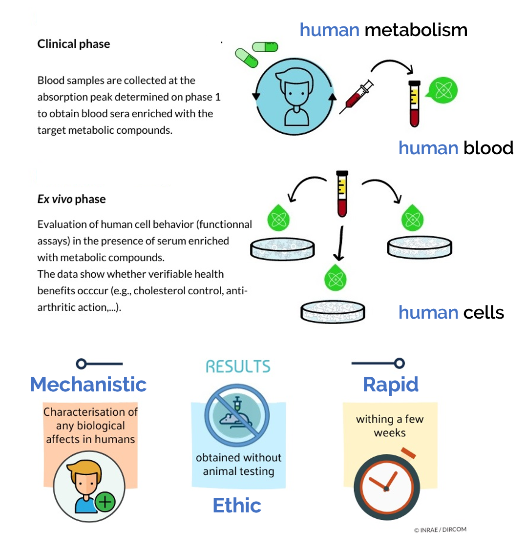
Abstract: Polyphenols are widely acknowledged for their health benefits, especially for the prevention of inflammatory and age-related diseases. We previously demonstrated that hydroxytyrosol (HT) and procyanidins (PCy), alone or in combination, drive preventive anti-osteoathritic effects in vivo. However, the lack of sufficient clinical evidences on the relationship between dietary phytochemicals and osteoarthritis remains. In this light, we investigated in humans the potential osteoarticular benefit of a grapeseed and olive extract (OPCO) characterized for its hydroxytyrosol (HT) and procyanidins (PCy) content. We first validated, in vitro, the anti-inflammatory and chondroprotective properties of the extract on primary cultured human articular chondrocytes stimulated by interleukin-1 beta (IL-1 β). The sparing effect involved a molecular mechanism dependent on the nuclear transcription factor-kappa B (NF-κB) pathway. To  confirm the clinical relevance of such a nutritional strategy,  we designed an innovative clinical approach taking into account the metabolites that are formed during the digestion process and that appear in circulation after the ingestion of the OPCO extract. Blood samples from volunteers were collected following ingestion, absorption, and metabolization of the extract and then were processed and applied on human primary chondrocyte cultures.    This original ex vivo methodology confirmed at a clinical level the chondroprotective properties previously observed in vitro and in vivo. Clinic’n’Cell ® has been registered as a trademark – clinicncell.com

Nutrients 2019, 11, 3071; doi:10.3390/nu11123071        www.mdpi.com/journal/nutrients

Keywords:  micronutrients;  osteoarthritis  (OA);  clinical  trials;  hydroxytyrosol;  procyanidins; cell biology

Clinic’n’Cell ® has been registered as a trademark – clinicncell.com

1.           Introduction

Osteoarthritis (OA) is a common inflammatory joint disease with a strong socioeconomic impact and a growing prevalence among the aging population [1,2]. From a pathophysiological point of view, OA is characterized by progressive cartilage loss, aberrant subchondral bone remodeling, osteophyte formation, and joint tissue inflammation [3,4]. This vicious cycle of inflammation and cartilage degradation is mainly driven by interleukin-1 beta (IL-1 β). IL-1 β stimulates the synthesis of nitric oxide (NO), prostaglandin E2 (PGE2), and matrix metalloproteinases (MMPs) [5,6]. Among these proteases, MMP-13 is overexpressed in OA and mediates cartilage type II collagen and aggrecan breakdown [7] feeding the amplification loop of inflammation.

Despite recent data further depicting OA mechanisms, OA management and treatments’ efficacy remain debated. Pharmacological approaches only alleviate inflammation and pain but do not slow down, stop, or reverse the progression of the cartilage degradation [8]. Moreover, most of these drug-based approaches are associated with side effects, including bleeding, ulceration, edema, or kidney failure. In this context, alternative strategies able to blunt the progression of the disease are required. The literature has recently highlighted the potential of nutritional compounds to target osteoarticular dysfunctions such as rheumatoid arthritis [9,10] or osteoarthritis [11–16]. In this light, we investigated in humans the potential osteoarticular benefit of a grapeseed and olive extract (OPCO) characterized for its hydroxytyrosol (HT) and procyanidins (PCy) content.

Hydroxytyrosol (HT) is a bioactive phenolic compound mainly found in olive fruit and oil. HT is known for its powerful antioxidant and anti-inflammatory properties including inhibition of PGE2 and NO production pathways [17,18]. Procyanidins (PCy) are active polyphenols found in many plants such as grape, pine bark, cocoa, and raspberry. PCy from grape seed extract has been reported to alleviate inflammation, to reduce NO and PGE2 both in vitro and in vivo, [19] and to blunt cartilage degradation during OA [17,20]. In a recently published paper, we demonstrated that hydroxytyrosol (HT) and procyanidins (PCy) used alone or in combination prevent post-traumatic osteoarthritis damages in both a destabilization of the medial meniscus (DMM) mouse model and an anterior cruciate ligament transection (ACLT) rabbit model and exhibit anti-IL-1 β activities in vitro and ex vivo in rabbit articular chondrocytes (RAC) cultures. However, the relationship between dietary phytochemicals and osteoarthritis at a clinical level remains unclear due to the lack of sufficient evidence [21].

Actually, despite this large body of in vitro data, “real life” cells within the body never interact with native nutrients. At a body level, cells within tissues deal with nutrient-derived metabolites [22] following oral administration. Along the digestive track, polyphenols undergo several chemical modifications before reaching the bloodstream [23,24].   Consistent with our previous in vitro and   in vivo data regarding HT and PCy influence on OA onset, we investigated the relevance of such     a nutritional strategy in humans. In this purpose, we designed a pioneering approach considering metabolism at the whole-body level to decipher whether and how different plant extracts enriched in HT and PCy may clinically benefit OA management. Clinic’n’Cell ® has been registered as a trademark – clinicncell.com

2.           Materials and Methods

2.1.        Ethics Clinical Trial

The investigations were carried out following the rules of the Declaration of Helsinki of 1975 (https:// www.wma.net/what-we-do/medical-ethics/declaration-of-helsinki/) revised in 2013. The human study was approved by the French Ethical Committee (Comité de Protection des Personnes (CPP17048/N◦ IDRCB: 2017-A02543-50) of Saint-Germain-en-Laye—Ile de France XI). No safety signals were reportedfollowing extract ingestion. The volunteers were informed on the objectives of the present study and on the potential risks of ingestion of polyphenols, such as diarrhea and abdominal pain.

2.2.        Human Study Design

A pool of 20 healthy men (20 to 30 years old; average: 24 years old; average BMI of 23.35 kg/m2; >50 kg; without treatment; and no distinction on ethnicity) volunteered for this study. They were tested for normal blood formulation and for renal and liver functions (aspartate aminotransferase (AST), alanine aminotransferase (ALT), gamma-glutamyltransferase (GGT), urea, and creatinine). Blood samples of all participants were obtained and collected in Vacutainer Ethylenediaminetetraacetic acid (EDTA)-containing tubes and serum-separating tubes for peripheral blood mononuclear cells (PBMCs) isolation and serum separation, respectively. Biological samples were prepared, aliquoted, and stored at the Centre de Ressources Biologiques (CRB)-Auvergne, a specialized laboratory that guarantees the quality of samples and compliance with regulatory and ethical obligations (certification according to the French standard NF S 96 900). The first step of the study aimed at determining the polyphenol absorption peak.  Five healthy volunteers who fasted for 12 h were given at once 3.2 g  of a grapeseed and olive extract (OPCO) characterized for its hydroxytyrosol (HT) and procyanidins (PCy) content. The dose was set according to validated preclinical data [23,24]. The OPCO extract was given under the form of 8 caps (400 mg each) with 200 mL of water. OPCO extract is a combination of grapeseed and olive extract with a guaranteed content in hydroxytyrosol (6%) and procyanidines (15%) and with a 90% total polyphenol content. Approximately 10 mL of venous blood was collected from the cubital vein, before and every 20 min after the ingestion for a total period of 240 min. Serum wasprepared from venous blood samples and stored at −80 ◦C until analysis. Clinic’n’Cell ® has been registered as a trademark – clinicncell.com

Polyphenol absorption profiles were quantified by ultra-high performance liquid chromatography (UPLC) coupled mass spectrometry. Once the absorption peak was determined, volunteers were called back for the collection of the enriched serum fraction. Twenty healthy volunteers who fasted for 12 h were given 3.2 g of OPCO extract under the form of 8 caps with 200 mL of water. Approximately 110 mL of venous blood was drawn from the cubital vein before the ingestion for the collection of a naïve serum. Then, at the maximum absorption peak, 100 mL of blood was drawn for enriched serum collection. Serum was stored at −80 ◦C until analysis.

2.3.        Phenolic Compounds Extraction from Serum

Sera (2.5 mL) from subjects fed as described above were mixed with 7.5 mL of methanol      100% for 2 min and ultrasonicated for 5 min. The mixture was centrifuged at 20,000 g for 10 min.  The supernatant was collected and evaporated to dryness using a SpeedVac Concentrator (Thermo Fisher Scientific, Illkirch, France). The dried extract was reconstituted in 200 µL of methanol/water (50:50 v/v). After centrifugation (20,000 g for 10 min), the supernatant was filtered through a polytetrafluoroethylene (PTFE) 0.22-µm filter (Millipore Corporation, Molsheim, France) and stored at −20 ◦C until use for ultra-high performance liquid chromatography-mass spectrometry (UPLC-MS) analysis.

2.4.        Ultra-High Performance Liquid Chromatography-Mass Spectrometry (UPLC-MS)

Phenolic compound analyses were carried out using a 1290 Infinity UPLC (Agilent Technologies, Les Ulis, France). The UPLC system was coupled to an Esquire 3000 plus mass spectrometer from Bruker Daltonics (Wissembourg, France). Five µL was injected into a column Zorbax SB-C18 (2.1 × 100 mm, 1.8 µm) (Agilent Technologies, Les Ulis, France). Two different solvents were used as a mobile phase: solvent A (water/formic acid 99.9:0.1, v/v) and solvent B (acetonitrile/formic acid 99.9:0.1, v/v), at a flow rate of 0.4 mL/min and a gradient as follows in solvent A: 0 min 1% B, 0.4 min 1% B, 2 min 10% B,     6 min 35% B, 7 min 50% B, 8.8 min 70% B, 10.8 min 92% B, 11 min 100% B, 12 min 100% B, 12.2 min 1% B, and 15.2 min 1% B. The MS/MS parameters were set as follows:  negative mode; capillary  tension +4000 V; nebulizer 40 psi; dry gas 10 L/min; dry temperature 365 ◦C; and scan range m/z 100 to 1400. Data were processed using HyStar 3.2 software (Bruker Daltonics, Wissembourg, France).

2.5.        OPC, PHO, and OPCO Solutions for In Vitro Experiments

Extracts were provided by Grap’Sud Union (Cruviers Lascours, France). They were characterized for their hydroxytyrosol, procyanidines, and total polyphenol contents as follows: (1) PHO extract (Olivex®, Grap’sud, Cruviers-Lascours, France), derived from olive, 6.5% of hydroxytyrosol/21% total polyphenol content;  (2) OPC extract (Exgrape®  SEED, Grap’sud, Cruviers-Lascours,   France), derived from grape seed, 30% of procyanidines/90% total polyphenol content; and (3) OPCO extract (Oleogrape®  SEED, Grap’sud, Cruviers-Lascours, France), derived from grapeseed and olive,   6.5% of hydroxytyrosol, 30% of procyanidines/90% total polyphenol content. Extracts were dissolved in Dulbecco’s Modified Eagle Medium high glucose (DMEM) (Thermo Fisher Scientific, Illkirch, France) to obtain a stock solution at 1 mg/mL. Solutions were sterilized by filtration through 0.22-µm membranes.

2.6.        Cell Culture

Human articular chondrocytes (HACs) were harvested from tibial plateau and femoral condyles following knee replacement surgery and isolated as previously described [25]. Only intact cartilage areas were kept and processed for chondrocytes isolation. Briefly, cartilage was sliced and chips were successively digested at 37 ◦C with 0.05% type IV-S hyaluronidase (750–3000 units/mg) (Sigma-Aldrich, Lyon, France) in Hank’s Balanced Sodium Salt (HBSS) (Life Technologies, Villebon-Sur-Yvette, France) for 10 min and then with 0.2% trypsin (≥9000 BAEE units/mg) (Sigma-Aldrich,  Lyon,  France) for   15 min and with 0.2% type II collagenase (125 units/mg) (Sigma-Aldrich, Lyon, France) for 30 min. Cartilage chips were then digested overnight at 37 ◦C in 0.03% type II collagenase in control medium (DMEM supplemented with 10% Fetal calf serum (FCS) (Pan-Biotech, Aidenbach, Germany) and 1% penicillin/streptomycin (Life Technologies, Villebon-Sur-Yvette, France). Cells were plated at passage 1 in F225 flasks at a density of 100,000 cells/cm2 and maintained at 37 ◦C in a humidified atmosphere of 5% CO2 in control medium (10% FCS, 1% P/S). At confluency, cells were subcultured for in vitro and ex vivo experiments. To analyze the effects of native polyphenols or their serum metabolites, cells were preincubated for 24 h in DMEM in the presence of native OPC, PHO, or OPCO extracts    at a concentration of 10 µg/mL (10% FCS and 1% P/S) or in the presence of 10% of human-enriched serum according to the Clinic’n’Cell protocol (DIRV INRA 18-00058) (1% P/S) prior an additional 24 h treatment with human recombinant IL-1 β (Millipore Corporation, Molsheim, France) at 1 ng/mL.

2.7.        Cell Proliferation

The in vitro cell proliferation was determined using MTS Assay Kit (Abcam 197010, Paris, France). The reduction of the MTS tetrazolium compound occurs in metabolically active cells. According to the supplier’s recommendations, the generation of the formazan dye was quantified by measuring the absorbance at 490–500 nm. The ex vivo cell proliferation was determined using an XTT-based method (Cell Proliferation Kit II, Sigma-Aldrich, Lyon, France) according to the supplier’s recommendations. Optical density was measured at 450 nm.

2.8.        NO, PGE2, and MMP-13 Quantification

Nitrate/Nitrite colorimetric assay and prostaglandin E2 Enzyme Immunoassay (EIA) kits were obtained from Cayman Chemical (Ann Arbor-MI, USA), and rabbit and human ELISA Kits for MMP-13 detection were purchased from Cloud-Clone Corp (Houston-TX, USA) and Abcam® (Paris, France) respectively. The NO, PGE2, and MMP-13 levels measurements were performed according to manufacturer’s instructions. For human serum, measurements were performed in quadruplicates for each sample of the twenty volunteers.

2.9.        Immunofluorescence Analyses

HACs were fixed in Phosphate Buffered Saline (PBS) (Life Technologies, Villebon-Sur-Yvette, France) containing 4% paraformaldehyde (PFA) (Electron Microscopy Science, Hatfield, PA, USA) and then permeabilized in PBS 0.2% Triton X-100 (Sigma-Aldrich, Lyon, France). Nonspecific binding sites were blocked and probed in PBS containing 1% Bovine Serum Albumin (BSA) (Sigma-Aldrich, Lyon, France).   Rabbit anti-NF-κB p65 primary antibodies (Cell Signaling Inc.,  Danvers-MA, USA)    were diluted 1/50 in PBS containing 1% BSA and were incubated overnight at 4 ◦C. Samples were incubated with Alexa Fluor® 488-conjugated secondary antibody (Life Technologies, Villebon-Sur-Yvette, France) diluted 1:1000 in 1% BSA in PBS for 3 h at room temperature. Cells nuclei were stained with Hoechst 33,258 Molecular Probes® (Life Technologies, Villebon-Sur-Yvette, France). HACs were counterstained with Phalloidin 594-conjugated at 1:50 in PBS for 72 h.

2.10.      Statistical Analysis

Each experiment was performed at least in triplicate. Results are expressed as mean ± SEM (standard error of the mean). Statistical analyses were carried out using ExcelStat Pro (Microsoft, Issy-les-Moulineaux, France). One-way ANOVA followed by Tukey’s test or T-test were performed. Groups with significant differences (P < 0.05) are indicated with different letters or * versus. Clinic’n’Cell ® has been registered as a trademark – clinicncell.com

3.           Results

3.1.        OPCO Extract Reduces IL-1ß-Induced Levels of NO, PGE2, and MMP-13 Production in Human Articular Chondrocytes (HACs)

We  first ascertained the effects of PHO, OPC, and OPCO extracts on cell viability.    HACs    were treated for 72 h  with  increasing  concentrations  of  the  different  extracts  (0,  0.5,  1,  5,  10,  50, and 100 µg/mL), and MTS activity was evaluated. When chondrocytes were cultured with actinomycin-D, a cell death inducer, MTS activity was significantly reduced by 80% as compared to its vehicle (DMSO –Dimethylsulfoxyde) (Figure 1). OPC and OPCO significantly decreased the MTS activity for concentration higher than 50 µg/mL in HAC. Consistently, the 10 µg/mL concentration for each extract was chosen for each extract and used for all subsequent experiments.

Figure 1. PHO, OPC, and OPCO effects on human articular chondrocytes proliferation rate. Human articular chondrocytes (HACs) were harvested from tibial plateau and femoral condyles following knee replacement surgery and isolated. Extracts were dissolved in Dulbecco’s Modified Eagle Medium high glucose (DMEM) (Thermo Fisher Scientific, Illkirch, France) to obtain a stock solution at 1 mg/mL. Solutions were sterilized by filtration through 0.22-µm membranes.      Cells were preincubated with PHO (A), OPC (B), or OPCO (C) extract solutions at different concentrations ranging from 0.5 to 100 µg/mL for 24 h and stimulated with IL-1 β (1 ng/mL) for additional 24 h. Effects of PHO, OPC, or OPCO on MTS activity were measured. PHO, OPC, or OPCO extracts are characterized for their content in hydroxytyrosol, procyanidins or both, respectively. Concentrations below 10 µg/mL have no significant impact on cell proliferation. NS: no significant difference compared to control condition (0 µg/mL).

To decipher the in vitro effects of PHO, OPC, and OPCO extracts on molecular mechanisms underlying OA, we evaluated whether extracts may modulate the expression levels of IL-1 β-responsive genes involved in inflammation and catabolic processes in cartilage. HACs were pre-treated with PHO, OPC, or OPCO extracts for 24 h and then stimulated or not with IL-1 β for additional 24 h. Levels of NO, PGE2, and MMP-13 were measured. As expected, IL-1 β treatment increased the production of markers involved in inflammation (NO +498% and PGE2 +4820%) and catabolism (MMP13 +427%) by human articular chondrocytes (Figure 2). This production was significantly limited in the presence of polyphenols from the different extracts. OPCO extract showed a greater limitation than both PHO and OPC on inflammatory marker production (NO from +498% to +104% of the control condition; PGE2 from +4820% to +15% of the control condition) (Figure 2A,B). Actually, OPCO totally suppressed the PGE2 production triggered by IL-1 β (Figure 2B). In contrast, although OPC, PHO, and OPCO extracts reduced MMP13 production (from +427% to +169% (PHO); from +427% to +275% (OPC); and from +427% to +234% (OPCO) of the control condition), there was no major difference between these three extracts (Figure 2C).

Figure 2. PHO, OPC, and OPCO effects on nitric oxide (NO), prostaglandin E2 (PGE2), and matrix metalloproteinase (MMP-13) production in human articular chondrocytes: Human articular chondrocytes (HACs) were harvested from tibial plateau and femoral condyles following knee replacement surgery and isolated. Extracts were dissolved in Dulbecco’s Modified Eagle Medium high glucose (DMEM) (Thermo Fisher Scientific, Illkirch, France). Solutions were sterilized by filtration through 0.22-µm membranes. Cells were preincubated with PHO, OPC, or OPCO extract solutions  at 10 µg/mL for 24 h and stimulated with IL-1β (1 ng/mL) for additional 24 h. Effects of PHO, OPC, and OPCO on NO (A), PGE2 (B), and MMP13 (C) release in culture media were measured. PHO, OPC, or OPCO extracts are characterized for their content in hydroxytyrosol, procyanidins, or both. Groups with significant differences (P < 0.05) are indicated with different letters (a, b, c, d, e and f). Clinic’n’Cell ® has been registered as a trademark – clinicncell.com

3.2.        OPCO Extract Decreases the IL-1 β-Mediated Activation of the NF-κB p65 Signaling Pathway

To determine the mechanism of action, we questioned whether IL-1 β-activated signaling pathway may be modulated by the presence of the polyphenols of the extracts. NF-κB is an essential transcriptional regulator of IL-1 β-activated inflammatory and catabolic mediators including NO, PGE2,  and MMP13.   After 24 h of pretreatment with the different extracts at 10 µg/mL followed    by an additional 24 h stimulation period with IL-1 β (1 ng/mL), the NF-κB p65 subunits were visualized by immunofluorescence assays in human articular chondrocytes (Figure 3). In untreated conditions, p65 NF-κB fragment mainly remained cytoplasmic with only a few human articular chondrocytes positive for nuclear p65 NF-κB fragment labelling (1.4%). In contrast, following IL-1 β incubation, the nuclear staining pattern for p65 NF-κB reached 74.7%, thus indicating a massive nuclear translocation. Pretreatment with PHO extract, OPC extract, or OPCO extract decreased nuclear staining of p65 NF-κB. According to fluorescence quantification, the limitations of the IL-1 β-induced nuclear translocation were −50.7%, −60.2%, and 54.8% for PHO, OPC, and OPCO respectively. Interestingly, p65 translocation was slightly but insignificantly upregulated by PHO, OPC, or OPCO extracts in the absence of IL-1 β stimulation. Clinic’n’Cell ® has been registered as a trademark – clinicncell.com

Figure 3. PHO, OPC, and OPCO effects on the IL-1 β-dependent nuclear transcription factor-kappa B (NF-κB) p65 translocation in human articular chondrocytes: Human articular chondrocytes (HACs) were harvested from tibial plateau and femoral condyles following knee replacement surgery and isolated. Extracts were dissolved in Dulbecco’s Modified Eagle Medium high glucose (DMEM) (Thermo Fisher Scientific, Illkirch, France). Solutions were sterilized by filtration through 0.22-µm membranes. Cells were preincubated with PHO, OPC, or OPCO extract solutions at 10 µg/mL for 24 h and stimulated with IL-1 β (1 ng/mL) for additional 24 h. Immunofluorescence assay for p65 subunit (A) was performed, and p65 positive cells in nucleus was counted (B). PHO, OPC, and OPCO extract solutions limited IL-1 β-induced p65 translocation to the nucleus. PHO, OPC, or OPCO extracts are characterized for their content in hydroxytyrosol, procyanidins, or both. Groups with significant differences (P < 0.05) are indicated with different letters (a, b and c). Clinic’n’Cell ® has been registered as a trademark – clinicncell.com

3.3.        Human-Enriched Sera Confirm the Chondroprotective Influence of Extracts / Clinic’n’Cell ® has been registered as a trademark – clinicncell.com

To get closer to a physiological context and to further investigate the anti-IL-1 β effects of OPCO extract, we validated the influence of the OPCO extract at a clinical level. Fasted volunteers received 3.2 g of OPCO extract. The digestion and absorption profile of the extract was monitored. Modulation of the polyphenol metabolite concentrations in human serum was measured by UPLC-MS. As shown in Figure 4, the cumulative area under curve (AUC) for all metabolites rapidly reached a peak at 100 min, corresponding to the maximum metabolite concentration time point. Consequently, enriched serum with OPCO metabolites was collected at 100 min post-ingestion.

Figure 4. Pharmacokinetics for metabolites in the serum from peripherical blood from 0 to 240 min after the ingestion of the OPCO extract: 5 healthy volunteers who fasted for 12 h were given 3.2 g of  a grapeseed and olive extract (OPCO) characterized for its hydroxytyrosol (HT) and procyanidins (PCy) content. The dose was set according to validated preclinical data. The OPCO extract was given under the form of 8 caps with 200 mL of water. Venous blood was collected from the cubital vein before and every 20 min after the ingestion for a total period of 240 min. Data are expressed as the mean values of 5 volunteers. Standard errors are depicted by vertical bars. The 100 min time point was chosen for enriched serum collection.

Then, we checked the influence of the different sera on cell proliferation and viability in primary human articular chondrocytes by measuring XTT-based activity. As expected, cell growth stopped  in serum free cultures (−18% and −42% after 24 h and 48 h, respectively, compared to 24 h of FCS 10%) while cells proliferated in the presence of FCS 10% (+31% between 24 and 48 h) (Figure 5A). Naïve or enriched human serum processed according to the Clinic’n’Cell methodology (DIRV#18-0058; see the Patents section) did not exert any cytotoxic effect on cell growth as compared to regular fetal calf serum and allowed cell proliferation similar to 10% FCS treatment (+35% between 24 and 48 h) (Figure 5B) [22].

To validate our cellular model and ex vivo methodology, we validated the influence of IL-1 β on cells in the presence of either FCS or human serum. As expected, IL-1β upregulated the production of NO, PGE2, and MMP13 in human primary articular chondrocytes. Upregulation occurred independently of the type of serum, FCS (Figure 6A–C) or human (Figure 6D–F), confirming the physiological relevance of both the model and the approach. Percentages of induction were similar in the presence of FCS or human serum reaching +159% and +121% for NO, +3982% and +2697% for PGE2, and +296% and +211% for MMP13, in the presence of FCS or human serum, respectively. Interestingly, as previously observed in vitro, the serum enrichment in OPCO extract metabolites significantly reduced the IL-1 β-induction of those three markers (−8.1% for NO, −50.7% for PGE2, and −5.8% for MMP13 production), validating the positive influence of OPCO on articular chondrocytes in humans. Clinic’n’Cell ® has been registered as a trademark – clinicncell.com

Figure 5. Effect of fetal calf serum and human serum enriched with OPCO metabolites on primary human articular chondrocytes proliferation: XTT activity. Human articular chondrocytes (HACs) were harvested from tibial plateau and femoral condyles following knee replacement surgery and isolated. Human primary chondrocytes were preincubated with serum from calf (A) or human origin (B) for 24 and 48 h. Primary human articular chondrocytes proliferation was measured using the XTT-based assay.   Cells proliferate without any negative impact of human serum or metabolite enrichment       (− absence; + presence). Groups with significant differences (P < 0.05) are indicated with different letters (a, b, c and d).

Figure 6. Effect of human serum enriched with OPCO metabolites on NO, PGE2, and MMP-13 production in primary human articular chondrocytes: Human articular chondrocytes (HACs) were harvested from tibial plateau and femoral condyles following knee replacement surgery and isolated. HACs were preincubated with fetal calf serum for 24 h and stimulated with IL-1 β (1 ng/mL) for additional 24 h (A–C). To analyze the effects of human serum enriched with metabolites, cells were preincubated for 24 h in DMEM in the presence of 10% of human-enriched serum according to the Clinic’n’Cell protocol (DIRV INRA 18-00058) (1% P/S) prior an additional 24 h treatment with human recombinant IL-1β (Millipore Corporation, Molsheim, France) at 1 ng/mL (D–F). NO (A,D), PGE2 (B,E), and MMP13 (C,F) release in culture media were measured. IL-1 β stimulated NO, PGE2, and MMP13 release independently of the origin of the serum (calf or human). The human serum enriched with OPCO metabolites significantly limited NO, PGE2, and MMP13 release.       * (P < 0.05);  ** (P < 0.01); *** (P < 0.001); **** (P < 0.0001). Clinic’n’Cell ® has been registered as a trademark – clinicncell.com

4.           Discussion

Clinic’n’Cell ® has been registered as a trademark – clinicncell.com

In this study, we demonstrated that PHO, OPC, or OPCO polyphenol extracts, characterized  for their content in hydroxytyrosol and/or procyanidins, exhibit anti-IL-1 β activities in human articular chondrocytes cultures both in vitro and ex vivo using an innovative human serum-enriched approach considering their metabolism. Despite outlying discrepancies between in vitro and ex vivo data, both approaches lead to the same conclusions and greatly match with the well-acknowledged anti-inflammatory capabilities of polyphenols [9]. For instance, in an experimental collagen-induced arthritis (CIA) mice arthritis model, extra-virgin olive-oil polyphenol extract containing hydroxytyrosol was proven to exert anti-inflammatory and joint-protective effects. When given at the doses of 100 and 200 mg/kg/day, these extracts reduced the levels of proinflammatory mediators (TNF-α, IFN-γ, IL-1 β, IL-6, IL-17 A, and PGE2) [26] and decreased serum IgG1 and IgG2a, cartilage olimeric matrix protein (COMP), and metalloproteinase-3 (MMP-3) [27].

Still, there is insufficient mechanism evidences on the relationship between dietary phytochemicals and osteoarthritis, especially in humans [21]. We investigated the impact of our extracts on the NF-κB pathway as a main signaling target in NO, PGE2, and MMP13 production [28].     PHO, OPC, and OPCO drastically reduced p65 nuclear translocation induced by the presence of IL-1 β. Our results nicely fit with literature data for rheumatoid arthritis models.   Hydroxytyrosol acetate (HTy-Ac)     is able to lower the activation of the Janus kinase-signal transducer and activator of transcription (JAK/STAT), mitogen-activated protein kinases (MAPKs), and nuclear transcription factor-kappa B (NF-κB) pathways and thus to downregulate the arthritic process in CIA mice [26,27]. In human fibroblast-like synoviocytes (FLSs) derived from arthritic tissues, resveratrol inhibits COX-2/PGE2 expression and reduces p65 phosphorylation [29].   Others signaling pathways may be involved      in polyphenol benefits but they were not investigated here. To date, resveratrol (stilbenes grape)  also inhibits TNF-alpha-induced MMP-3 production in human rheumatoid arthritis fibroblast-like synoviocytes via a modulation of the PI3kinase/Akt pathway [30].

Polyphenol’s  benefits  on  health  may  be  potentialized  by  other  nutrients.  For  instance,  the combination of curcuminoid extract,  hydrolyzed collagen,  and green tea extract was proven    to be significantly more efficient in inhibiting interleukin-1 β-stimulated matrix metalloproteinase-3 expression than curcuminoid extract alone in normal bovine chondrocytes and osteoarthritic human chondrocytes cultures [31,32]. The same authors described a trended reduction of pain in an OA model when dogs were fed with the corresponding mixture for 3 months [33]. Remarkably, a synergistic effect greater than the sum of the effects observed separately for each compound has even been described for fisetin and docosahexaenoic acid (DHA) on bone with an inhibition of osteoclastogenesis when compounds were mixed [34,35]. OPCO gathers part of the OPC and PHO compositions regarding HT and PCy content. Thus, we wondered if OPCO composition may result in synergistic or cumulative benefits. Unfortunately, we found no synergy but, somehow, found a few cumulative effects that depend on the target. Actually, additional effects were observed for NO and PGE2 production in vitro in HAC but not for MMP13. This observation suggests that, in contrast to MMP13, PHO (HT) and OPC (PCy) extracts may influence NO and PGE2 production through different pathways. Both PHO (HT) and OPC (PCy) almost completely blocked p65 translocation, thus providing insight regarding the absence of a cumulative effect of the OPCO (HT/PCy) on the NF-κB pathway. Thus, while NF-κB blocking by PHO (HT) or OPC (PCy) may at least partly explain the absence of a cumulative effect by OPCO (HT/PCy) on MMP13, it does not fully address the mechanism for cumulative effect on PGE2 and NO, supporting the involvement of other signaling pathways. absorption, and transformation through the digestive track. Therefore, to improve the relevance of our

We previously published the effect of both HT and PCy in a rabbit model of OA. According to the protocol, the quantity of polyphenols (including HT and PCy) delivered at once and given by force-feeding was set to 100 mg/kg of body weight [23,24]. According to metabolic weight calculation, this dose corresponds to 45 mg of polyphenols per kilogram of body weight in humans or a single dose of 3 g of polyphenols. Thus, volunteers were given orally 3.2 g of the OPCO extract containing 95% of polyphenols including 15% of PCy and 6% of HT. The recommendation for the polyphenol daily intake surrounds 1 g [36]. Although slightly higher, the dose delivered remains in a nutritional range and, overall, ten times lower than the dose delivered to our mouse model of OA showing positive impact of the extract on the AO score with no side effects [23,24]. the approach (+159% and +121% for NO; +3982% and +2697% for PGE2; and +296% and +211% depending on HAC batch. Indeed, differences between Figures 2 and 5 were related to donors but, beside the seemingly discrepancy observed, variations remained in the same range (+159% and +500% for NO; +3982% and +4800% for PGE2; and +296% and +427% for MMP13 depending on HAC batches).

Although, it had slight but no significant effect on transcripts (Figure A1), the serum enrichment in OPCO extract metabolites significantly limited the IL-1 β-induction of those three markers (−8.1% for NO, −50.7% for PGE2, and −5.8% for MMP13 production). These data further confirm at a clinical level the chondroprotective role of the extract that we observed in vitro upon pro-inflammatory stress. Moreover, these ex vivo results perfectly match with literature data from other clinical trials regarding anti-inflammatory and antioxidant capabilities of “polyphenol strategies”. To date, a reconstituted freeze-dried strawberry beverage (50 g/day) given to obese adults with radiographic evidence of knee OA significantly lowered serum biomarkers of inflammation including (IL)-6, IL-1 β, and cartilage degradation mediators including matrix metalloproteinase (MMP)-3 [37]. Forty g of freeze-dried blueberry powder given daily for four months to adults with symptomatic knee OA significantly decreased (WOMAC, Western Ontario and McMaster Universities Osteoarthritis Index) total score and subgroups, including pain, stiffness, and difficulty to perform daily activities [38]. Pomegranate juice decreases serum levels of matrix metalloproteinase (MMP)-13 and improves antioxidant status (glutathione peroxidase) in patients with knee OA [39]. Remarkably, pomegranates and blueberries are well-acknowledged sources of both PCy and anthocyanins [40,41]. Curcuminoids in combination with hydroxytyrosol attenuates systemic oxidative stress in patients with mild-to-moderate primary knee osteoarthritis when given at the dose of 1500 mg/day for a period of 6 weeks [42].

Among other classes of polyphenols, resveratrol has attracted much attention. Used as an adjuvant with meloxicam, resveratrol (500 mg/day) significantly improves pain, knee functions, and WOMAC scores with no side effects in patients with knee osteoarthritis [43].  Given twice daily at the dose     of 75 mg for 14 weeks, it reduces chronic pain in healthy postmenopausal women with age-related osteoarthritis [44]. Finally, in patients with rheumatoid arthritis, activity score assessment for 28 joints was significantly lowered when their conventional treatment was supplemented with a daily capsule of 1 g of resveratrol for 3 months [45].

Although minor compared to the in vitro observations, the significant limitation of NO, PGE2, and MMP13 release reported by our ex vivo methodology strongly supports the relevance of a “realistic” polyphenol strategy for the management of chronic OA. The rationale of our pioneering clinical methodology was recently reinforced as polyphenol metabolites were found to distribute into the serum but, more interestingly, into the synovial fluid of patients with osteoarthritis [46]. Remarkably, our conclusions strictly parallel with clinical trials that were run for several months and needed      to recruit hundreds of volunteers to evidence the chondroprotective effect and to reach a statistical significance.      In this context, although our approach would not substitute a “regular” clinical trial, this innovative clinical approach may have helped the authors to rapidly explore human data before moving into a huge clinical investment [22]. Clinic’n’Cell ® has been registered as a trademark – clinicncell.com

5.           Conclusions

From a global perspective, our results correlate not only with literature data on the positive role of polyphenols but also, most notably, with those from olive and grape origin on cartilage. Using a pioneering clinical screening approach, we further support the articular sparing ability of HT- and PCy-enriched extracts and the importance of the NF-κB pathway on the mode of action   of polyphenols on cartilage metabolism. Accordingly, grapeseed and olive extracts characterized for their HT and PCy content stand as a relevant nutritional opportunity for advanced strategies to   manage osteoarticular health conditions. Clinic’n’Cell ® has been registered as a trademark – clinicncell.com

6.           Patents

 The human ex vivo methodology used in this study has been registered as a written   invention disclosure by the French National Institute for Agronomic Research (INRA) (DIRV#18-0058). Clinic’n’Cell® has been registered as a mark [22]. Clinic’n’Cell ® has been registered as a trademark – clinicncell.com

Author Contributions: Conceptualization, Y.W., F.W., C.S.V., and  L.B.-W.;  methodology,  Y.W.  and  F.W.; formal analysis, E.M., S.K., T.R., J.V., R.H.-O., F.W.,  H.G.,  and  J.G.;  clinical  investigation,  A.B.,  N.M.,  J.B., S.D., and Y.W.; writing—original draft preparation, Y.W.; writing—review and editing, F.W., A.B., N.M., and C.S.V.; project administration, N.U., L.B.-W., L.B., and Y.W.; funding acquisition, N.U., F.W., and Y.W.

Funding: We thank Clermont-Auvergne Métropole for funding Fabien Wauquier and Line Boutin-Wittrant (Grant Clermont Création Innovation) and Université Clermont Auvergne for hosting them.

Acknowledgments: We thank all the CRNH team, Françoise Laporte, Hélène Parrot, Véronique Pidou, Dominique Provenchère, and Amandine Prulière for their help and relevant clinical follow-up. Parts of the TOC graphic used Servier Medical Art according to the licence Creative Commons Attribution 3.0 France.

Conflicts of Interest: Fabien Wauquier, Elsa Mevel, Stephanie Krisa, Tristan Richard, Josep Valls, Ruth Hornedo Ortega, Henri Granel, Juliette Berger, Stéphane Descamps, Jérôme Guicheux, Laurent Beck, Claire Vinatier, Nathalie Meunier, Adeline Blot, and Yohann Wittrant have no conflict of interest to declare. Nelly Urban works for Inosud and provided the extracts.

Appendix A

Figure A1. Effect of human serum enriched with OPCO metabolites on MMP-13 and PTGS2 mRNA expression levels in primary human articular chondrocytes: Human articular chondrocytes were preincubated with human serum enriched with OPCO metabolites for 24 h and stimulated with IL-1 β (1 ng/mL) for additional 24 h (- absence; + presence).  MMP-13 and PTGS2 mRNA expression  levels in primary human articular chondrocytes was measured by RT-PCR (PowerUp SYBRgreen, Applied Biosystems). β-Actine was used as a housekeeping gene. Primers were designed as follows: PTGS2-F: CTT CAC GCA TCA GTT TTT CAA G; PTGS2-R: TCA CCG TAA ATA TGA TTT AAG TCC AC; MMP13-F: CCA GTC TCC GAG GAG AAA CA; MMP13-R: AAA AAC AGC TCC GCA TCA AC; ACTB-F: ATT GGC AAT GAG CGG TTC; and ACTB-R: GGA TGC CAC AGG ACT CCA.

The incubation with IL-1 β (1 ng/mL) was set as the control condition to enlighten the effect of the human serum enriched with OPCO metabolites. Despite a trend, there was no significant impact of the enrichment. Groups with significant differences (P < 0.05) are indicated with different letters (a and b).

References

1.           Nuesch, E.; Dieppe, P.; Reichenbach, S.; Williams, S.; Iff, S.; Juni, P. All cause and disease specific mortality in patients with knee or hip osteoarthritis: Population based cohort study. BMJ 2011, 342, d1165. [CrossRef] [PubMed]

2.           Bruyere, O.; Cooper, C.; Al-Daghri, N.M.; Dennison, E.M.; Rizzoli, R.; Reginster, J.Y. Inappropriate claims from non-equivalent medications in osteoarthritis:  A position paper endorsed by the European Society  for Clinical and Economic Aspects of Osteoporosis, Osteoarthritis and Musculoskeletal Diseases (ESCEO). Aging Clin. Exp. Res. 2018, 30, 111–117. [CrossRef] [PubMed]

3.           Bijlsma, J.W.;  Berenbaum, F.;  Lafeber, F.P.  Osteoarthritis:  An update with relevance for clinical    practice.

Lancet 2011, 377, 2115–2126. [CrossRef]

4.           Loeser, R.F.; Goldring, S.R.; Scanzello, C.R.; Goldring, M.B. Osteoarthritis: A disease of the joint as an organ.

Arthritis Rheum. 2012, 64, 1697–1707. [CrossRef] [PubMed]

5.           Karsdal, M.A.; Madsen, S.H.; Christiansen,  C.;  Henriksen,  K.;  Fosang,  A.J.;  Sondergaard,  B.C.  Cartilage degradation is fully reversible in the presence of aggrecanase but not matrix metalloproteinase activity. Arthritis Res. Ther. 2008, 10, R63. [CrossRef]  [PubMed]

6.           Lim, N.H.; Kashiwagi, M.; Visse, R.;  Jones, J.;  Enghild, J.J.;  Brew,  K.;  Nagase, H. Reactive-site mutants  of N-TIMP-3 that selectively inhibit ADAMTS-4 and ADAMTS-5: Biological and structural implications. Biochem. J. 2010, 431, 113–122. [CrossRef]

7.           Deberg, M.; Labasse, A.; Christgau, S.; Cloos, P.; Bang Henriksen, D.; Chapelle, J.P.; Zegels, B.; Reginster, J.Y.; Henrotin, Y. New serum biochemical markers (Coll 2-1 and Coll 2-1 NO2) for studying oxidative-related type II collagen network degradation in patients with osteoarthritis and rheumatoid arthritis. Osteoarthr. Cartil. 2005, 13, 258–265. [CrossRef]

8.           Clouet, J.; Vinatier, C.; Merceron, C.; Pot-vaucel, M.; Maugars, Y.; Weiss, P.; Grimandi, G.; Guicheux, J. From osteoarthritis treatments to future regenerative therapies for cartilage. Drug Discov. Today 2009, 14, 913–925. [CrossRef]

9.           Sung, S.; Kwon, D.; Um, E.; Kim, B. Could Polyphenols Help in the Control of Rheumatoid Arthritis?

Molecules 2019, 24, 1589. [CrossRef]

10.         Bang, J.S.; Oh, D.H.; Choi, H.M.; Sur, B.J.; Lim, S.J.; Kim, J.Y.; Yang, H.I.; Yoo, M.C.; Hahm, D.H.; Kim, K.S. Anti-inflammatory and antiarthritic effects of piperine in human interleukin 1beta-stimulated fibroblast-like synoviocytes and in rat arthritis models. Arthritis Res. Ther. 2009, 11, R49. [CrossRef]

11.         Chen, W.P.; Hu, P.F.; Bao, J.P.; Wu, L.D. Morin exerts antiosteoarthritic properties: An in vitro and in vivo study. Exp. Biol. Med. 2012, 237, 380–386. [CrossRef]  [PubMed]

12.         Chen, W.P.; Tang, J.L.; Bao, J.P.; Hu, P.F.; Shi, Z.L.; Wu, L.D. Anti-arthritic effects of chlorogenic acid in interleukin-1beta-induced rabbit chondrocytes and a rabbit osteoarthritis model. Int. Immunopharmacol. 2011, 11, 23–28. [CrossRef] [PubMed]

13.         Chen, W.P.; Wang, Y.L.; Tang, J.L.; Hu, P.F.; Bao, J.P.; Wu, L.D. Morin inhibits interleukin-1beta-induced nitric oxide and prostaglandin E2 production in human chondrocytes. Int. Immunopharmacol. 2012, 12, 447–452. [CrossRef] [PubMed]

14.         Haseeb, A.; Chen, D.; Haqqi, T.M. Delphinidin inhibits IL-1beta-induced activation of NF-kappaB by modulating the phosphorylation of IRAK-1(Ser376) in human articular chondrocytes. Rheumatology 2013, 52, 998–1008. [CrossRef] [PubMed]

15.         Phitak, T.; Pothacharoen, P.; Settakorn, J.; Poompimol, W.; Caterson, B.; Kongtawelert, P. Chondroprotective and anti-inflammatory effects of sesamin. Phytochemistry 2012, 80, 77–88. [CrossRef] [PubMed]

16.         Wang, J.; Gao, J.S.; Chen, J.W.; Li, F.; Tian, J. Effect of resveratrol on cartilage protection and apoptosis inhibition in experimental osteoarthritis of rabbit. Rheumatol. Int. 2012, 32, 1541–1548. [CrossRef]

17.         Aini, H.; Ochi, H.; Iwata, M.; Okawa, A.; Koga, D.; Okazaki, M.; Sano, A.; Asou, Y. Procyanidin B3 prevents articular cartilage degeneration and heterotopic cartilage formation in a mouse surgical osteoarthritis model. PLoS ONE 2012, 7, e37728. [CrossRef]

18.         Bascoul-Colombo, C.; Garaiova, I.; Plummer, S.F.; Harwood, J.L.; Caterson,  B.;  Hughes,  C.E.  Glucosamine Hydrochloride but Not Chondroitin Sulfate Prevents Cartilage Degradation and Inflammation Induced by Interleukin-1alpha in Bovine Cartilage Explants. Cartilage 2016, 7, 70–81. [CrossRef]

19.         Pallares, V.; Fernandez-Iglesias, A.; Cedo, L.; Castell-Auvi, A.; Pinent, M.; Ardevol, A.; Salvado, M.J.; Garcia-Vallve, S.; Blay, M. Grape seed procyanidin extract reduces the endotoxic effects induced by lipopolysaccharide in rats. Free Radic. Biol. Med. 2013, 60, 107–114.  [CrossRef]

20.         He, L.; Mu, C.; Shi, J.; Zhang, Q.; Shi, B.; Lin, W. Modification of collagen with a natural cross-linker, procyanidin. Int. J. Biol. Macromol. 2011, 48, 354–359.  [CrossRef]

21.         Guan, V.X.; Mobasheri, A.; Probst, Y.C. A systematic review of osteoarthritis prevention and management with dietary phytochemicals from foods. Maturitas 2019, 122, 35–43. [CrossRef] [PubMed]

22.         Wauquier, F.; Daneault, A.; Granel, H.; Prawitt, J.; Fabien Soule, V.; Berger, J.; Pereira, B.; Guicheux, J.; Rochefort, G.Y.; Meunier, N.; et al. Human Enriched Serum Following Hydrolysed Collagen Absorption Modulates Bone Cell Activity: From Bedside to Bench and Vice Versa. Nutrients 2019, 11, 1249. [CrossRef] [PubMed]

23.         Mevel, E.; Merceron, C.; Vinatier, C.; Krisa, S.; Richard, T.; Masson, M.; Lesoeur, J.; Hivernaud, V.; Gauthier, O.; Abadie, J.; et al. Olive and grape seed extract prevents post-traumatic osteoarthritis damages and exhibits in vitro anti IL-1beta activities before and after oral consumption. Sci. Rep. 2016, 6, 33527. [CrossRef] [PubMed]

24.         Mevel, E.; Monfoulet, L.E.; Merceron, C.; Coxam, V.; Wittrant, Y.; Beck, L.; Guicheux, J. Nutraceuticals in joint health: Animal models as instrumental tools. Drug Discov. Today 2014, 19, 1649–1658. [CrossRef] [PubMed]

25.         Vinatier, C.; Magne, D.; Weiss, P.; Trojani, C.; Rochet, N.; Carle, G.F.; Vignes-Colombeix, C.; Chadjichristos, C.; Galera, P.; Daculsi, G.; et al. A silanized hydroxypropyl methylcellulose hydrogel for the three-dimensional culture of chondrocytes. Biomaterials 2005, 26, 6643–6651. [CrossRef] [PubMed]

26.         Rosillo, M.A.; Alcaraz, M.J.; Sanchez-Hidalgo, M.; Fernandez-Bolanos, J.G.; Alarcon-de-la-Lastra, C.; Ferrandiz, M.L. Anti-inflammatory and joint protective effects of extra-virgin olive-oil polyphenol extract in experimental arthritis. J. Nutr. Biochem. 2014, 25, 1275–1281. [CrossRef]

27.         Rosillo, M.A.; Sanchez-Hidalgo, M.; Gonzalez-Benjumea, A.; Fernandez-Bolanos, J.G.; Lubberts, E.; Alarcon-de-la-Lastra, C. Preventive effects of dietary hydroxytyrosol acetate, an extra virgin olive oil polyphenol in murine collagen-induced arthritis. Mol. Nutr. Food Res. 2015, 59, 2537–2546.  [CrossRef]

28.         Chen, C.; Zhang, C.; Cai, L.; Xie, H.; Hu, W.; Wang, T.; Lu, D.; Chen, H. Baicalin suppresses IL-1beta-induced expression of inflammatory cytokines via blocking NF-kappaB in human osteoarthritis chondrocytes and shows protective effect in mice osteoarthritis models. Int. Immunopharmacol. 2017, 52, 218–226. [CrossRef]

29.         Tsai, M.H.; Hsu, L.F.; Lee, C.W.; Chiang, Y.C.; Lee, M.H.; How, J.M.; Wu, C.M.; Huang, C.L.; Lee, I.T. Resveratrol inhibits urban particulate matter-induced COX-2/PGE2 release in human fibroblast-like synoviocytes via the inhibition of activation of NADPH oxidase/ROS/NF-kappaB. Int. J. Biochem. Cell Biol. 2017, 88, 113–123. [CrossRef]

30.         Tian, J.; Chen,  J.W.;  Gao,  J.S.;  Li,  L.;  Xie,  X.  Resveratrol  inhibits  TNF-alpha-induced  IL-1beta,  MMP-3 production in human rheumatoid arthritis fibroblast-like synoviocytes via modulation of PI3kinase/Akt pathway. Rheumatol. Int. 2013, 33, 1829–1835.  [CrossRef]

31.         Comblain, F.; Sanchez, C.; Lesponne, I.; Balligand, M.; Serisier, S.; Henrotin, Y. Curcuminoids extract, hydrolyzed collagen and green tea extract synergically inhibit inflammatory and catabolic mediator’s synthesis by normal bovine and osteoarthritic human chondrocytes in monolayer. PLoS ONE 2015, 10, e0121654. [CrossRef] [PubMed]

32.         Comblain, F.; Dubuc, J.E.; Lambert, C.; Sanchez, C.; Lesponne, I.; Serisier, S.; Henrotin, Y. Identification of Targets of a New Nutritional Mixture for Osteoarthritis Management Composed by Curcuminoids Extract, Hydrolyzed Collagen and Green Tea Extract. PLoS ONE 2016, 11, e0156902. [CrossRef] [PubMed]

33.         Comblain, F.; Barthelemy, N.; Lefebvre, M.; Schwartz, C.; Lesponne, I.; Serisier, S.; Feugier, A.; Balligand, M.; Henrotin, Y. A randomized, double-blind, prospective, placebo-controlled study of the efficacy of a diet supplemented with curcuminoids extract, hydrolyzed collagen and green tea extract in owner’s dogs with osteoarthritis. BMC Vet. Res. 2017, 13, 395. [CrossRef]  [PubMed]

34.         Leotoing, L.; Coxam, V.; Wittrant, Y. Use of a Combination of Two Compounds for the Treatment and/or Prevention of Bone Disorders; European Patent Office: Munich, Germany, 2014; p. EP2999467A1.

35.         Leotoing, L.; Wauquier, F.; Guicheux, J.; Miot-Noirault, E.; Wittrant, Y.; Coxam, V. The polyphenol fisetin protects bone by repressing NF-kappaB and MKP-1-dependent signaling pathways in osteoclasts. PLoS ONE 2013, 8, e68388. [CrossRef]

36.         Williamson, G.; Holst, B. Dietary reference intake (DRI) value for dietary polyphenols: Are we heading in the right direction? Br. J. Nutr. 2008, 99, S55–S58.  [CrossRef]

37.         Schell, J.; Scofield, R.H.; Barrett, J.R.; Kurien, B.T.; Betts, N.; Lyons, T.J.;  Zhao,  Y.D.;  Basu,  A.  Strawberries Improve Pain and Inflammation in Obese Adults with Radiographic Evidence of Knee Osteoarthritis. Nutrients 2017, 9, 949. [CrossRef]

38.         Du, C.; Smith, A.; Avalos, M.; South, S.; Crabtree, K.; Wang, W.; Kwon, Y.H.; Vijayagopal, P.; Juma, S. Blueberries Improve Pain, Gait Performance, and Inflammation in Individuals with Symptomatic Knee Osteoarthritis. Nutrients 2019, 11, 290. [CrossRef]

39.         Ghoochani, N.; Karandish, M.; Mowla, K.; Haghighizadeh, M.H.; Jalali, M.T. The effect of pomegranate juice on clinical signs, matrix metalloproteinases and antioxidant status in patients with knee osteoarthritis. J. Sci. Food Agric. 2016, 96, 4377–4381. [CrossRef]

40.         Spilmont, M.; Leotoing, L.; Davicco, M.J.; Lebecque, P.; Mercier, S.; Miot-Noirault, E.; Pilet, P.; Rios, L.; Wittrant, Y.; Coxam, V. Pomegranate and its derivatives can improve bone health through decreased inflammation and oxidative stress in an animal model of postmenopausal osteoporosis. Eur. J. Nutr. 2014, 53, 1155–1164. [CrossRef]

41.         Davicco, M.J.; Wittrant, Y.; Coxam, V. Berries, their micronutrients and bone health. Curr. Opin. Clin. Nutr. Metab. Care 2016, 19, 453–457. [CrossRef] [PubMed]

42.         Panahi, Y.; Alishiri, G.H.; Parvin, S.; Sahebkar, A. Mitigation of Systemic Oxidative Stress by Curcuminoids in Osteoarthritis: Results of a Randomized Controlled Trial. J. Diet. Suppl. 2016, 13, 209–220. [CrossRef] [PubMed]

43.         Hussain, S.A.; Marouf, B.H.; Ali, Z.S.; Ahmmad, R.S. Efficacy and safety of co-administration of resveratrol with meloxicam in patients with knee osteoarthritis: A pilot interventional study. Clin. Interv. Aging 2018, 13, 1621–1630. [CrossRef] [PubMed]

44.         Wong, R.H.X.; Evans, H.M.; Howe, P.R.C. Resveratrol supplementation reduces pain experience by postmenopausal women. Menopause 2017, 24, 916–922. [CrossRef] [PubMed]

45.         Khojah, H.M.; Ahmed, S.; Abdel-Rahman, M.S.; Elhakeim, E.H. Resveratrol as an effective adjuvant therapy in the management of rheumatoid arthritis: A clinical study. Clin. Rheumatol. 2018, 37, 2035–2042. [CrossRef] [PubMed]

46.         Mulek, M.; Seefried, L.; Genest, F.; Hogger, P. Distribution of Constituents and Metabolites of Maritime Pine Bark Extract (Pycnogenol((R))) into Serum, Blood Cells, and Synovial Fluid of Patients with Severe Osteoarthritis: A Randomized Controlled Trial. Nutrients 2017, 9, 443. [CrossRef] © 2019 by the authors. Licensee MDPI, Basel, Switzerland. This article is an open access article distributed under the terms and conditions of the Creative Commons Attribution (CC BY) license (http://creativecommons.org/ licenses/by/4.0/).

1st publication in May 2019 : ClinicnCell concept validation

Human Enriched Serum Following Hydrolysed Collagen Absorption Modulates Bone Cell Activity: from Bedside to Bench and Vice Versa / ClinicnCell

Clinic’n’Cell ® has been registered as a trademark – clinicncell.com

https://www.ncbi.nlm.nih.gov/pmc/articles/PMC6627680/

https://www.ncbi.nlm.nih.gov/pmc/articles/PMC6627680/pdf/nutrients-11-01249.pdf

https://www.mdpi.com/2072-6643/11/6/1249/htm

Fabien Wauquier 1,2, Audrey Daneault 1,2, Henri Granel 1,2, Janne Prawitt 3, Véronique Fabien Soulé 4, Juliette Berger 5, Bruno Pereira 6 , Jérôme Guicheux 7,8,9, Gael Y. Rochefort 7, Nathalie Meunier 10, Adeline Blot 10  and Yohann Wittrant 1,2,*

1             INRA, UMR 1019, UNH, CRNH Auvergne, F-63009 Clermont-Ferrand, France; Fabien_Wauquier@gmx.fr (F.W.) from Clinic’n’Cell / ClinicnCell ; audrey.daneault@gmail.com (A.D.); henri.granel@inra.fr (H.G.)

2             Unité de Nutrition Humaine, Clermont Université, Université d’Auvergne, BP 10448 F-63000 Clermont-Ferrand, France

3             Rousselot BVBA, Meulestedekaai 81, 9000 Gent, Belgium; janne.prawitt@rousselot.com

4             Rousselot SAS, 4 rue de l’abreuvoir, 92400 Courbevoie, France; vfabiensoule@syndifrais.com

5             CRB Auvergne, Hématologie Biologique, Equipe d’Accueil 7453 CHELTER, Centre Hospitalier Universitaire Estaing, 1 place Lucie et Raymond Aubrac, CEDEX 1, F-63003 Clermont-Ferrand, France; jberger@chu-clermontferrand.fr

6             Biostatistics Unit (DRCI), University hospital Clermont-Ferrand, 58 rue Montalembert,

63000 Clermont-Ferrand, France; bpereira@chu-clermontferrand.fr

7             Inserm, UMR 1229, RMeS, Regenerative Medicine and Skeleton, Université de Nantes, ONIRIS, 44000 Nantes, France; jerome.guicheux@inserm.fr (J.G.); gael.rochefort@gmail.com (G.Y.R.)

8             UFR Odontologie, Université de Nantes, 44000 Nantes, France

9             CHU Nantes, PHU4 OTONN, 44000 Nantes, France

10          Chu Clermont-Ferrand, Centre De Recherche En Nutrition Humaine Auvergne, 58 rue Montalembert, 63000 Clermont-Ferrand, France; nmeunier@chu-clermontferrand.fr (N.M.); ablot@chu-clermontferrand.fr (A.B.)

Clinic’n’Cell®has been registered as a trademark (Clinic’n’Cell / ClinicnCell).

*             Correspondence: yohann.wittrant@inra.fr; Tel.: +33-(0)473624784

Received: 24 April 2019; Accepted: 28 May 2019; Published: 31 May 2019

Abstract: Collagen proteins are crucial components of the bone matrix. Since collagen-derived products are widely used in the food and supplement industry, one may raise the question whether collagen-enriched diets can provide benefits for the skeleton. In this study, we designed an innovative approach to investigate this question taking into account the metabolites that are formed by the digestive tract and appear in the circulation after ingestion of hydrolysed collagen. In this innovative format we called Clinic’n’Cell or ClinicnCell (clinicncell.com), blood samples were collected in clinical and pre-clinical trials following ingestion and absorption of hydrolysed collagen and were processed and applied on bone-related primary cell cultures. This original ex vivo methodology revealed that hydrolysed collagen-enriched serum had a direct impact on the behaviour of cells from both human and mouse origin that was not observed with controls (bovine serum albumin or hydrolysed casein-enriched serum). These ex vivo findings were fully in line with in vivo results obtained from a mouse model of post-menopausal osteoporosis. A significant reduction of bone loss was observed in mice supplemented with hydrolysed collagen compared to a control protein. Both the modulation of osteoblast and osteoclast activity observed upon incubation with human or mouse serum ex vivo and the attenuation of bone loss in vivo, clearly indicates that the benefits of hydrolysed collagen for osteoporosis prevention go beyond the effect of a simple protein supplementation.

Keywords:  metabolites;  bone;  hydrolysed  collagen;  nutrition;  osteoporosis;  absorption; collagen peptides

Clinic’n’Cell®has been registered as a trademark (Clinic’n’Cell / ClinicnCell).

Nutrients 2019, 11, 1249; doi:10.3390/nu11061249        www.mdpi.com/journal/nutrients

https://clinicncell.com/

1.           Introduction

Osteoporosis is a major cause of morbidity and disability and is considered as an important contributor to medical care costs worldwide. Several treatment options are available, but concerns about possible side effects including an increased risk for cancer and cardiovascular disease have been raised [1]. Thus, prophylaxis strategies and early prevention by nutritional interventions may offer relevant alternatives [2–4]. The primary aim of a nutritional strategy for the prevention of osteoporosis is to provide persons at risk with a sufficient and bioavailable amount of nutrients that favours bone growth and remodelling [5]. Proteins play a major role in skeleton metabolism by providing building blocks and by exerting specific regulatory functions. Thus, protein-based supplements are promising candidates to maintain bone health during aging.

Collagen is the major structural element in the extracellular matrix of all connective tissues, including bone, and represents the most abundant protein in mammals accounting for 30% of the total protein mass in the body and 80% in the skeleton (mostly type I) [6]. In bone, collagen plays an important role in the force transmission and tissue structure maintenance [7,8]. Collagen comprises three polypeptide strands (alpha-chains) which form a unique triple-helical structure. Each strand is built by the repeating amino acid sequence Gly-X-Y, with every third amino acid being a glycine and with X and Y being mainly proline (Pro) and hydroxyproline (Hyp).

Collagen and its derivatives (gelatin and hydrolysed collagen) can be extracted from bone or skin and are widely used in the food, cosmetic and pharmaceutical industry. Hydrolysed collagen (HC), produced by enzymatic hydrolysis from gelatin, has been reported to improve joint pain and function in patients suffering from osteoarthritis [9–14], but there is a clear lack of clinical data for direct effects of HC on the skeleton. In humans, HC have mainly been used in association with other compounds such as calcium-collagen chelate, vitamin D [3,15] or curcuminoids [16] to investigate benefits on bone health, and the Group for the Respect of Ethics and Excellence in Science has comprehensively outlined that further studies are warranted to strengthen the scientific evidence, including underlying pathways, for HC effects on the bone [17].

Consistent with the growing demand for innovative tools of high physiological relevance, that can assess the efficacy of nutritional interventions, we have designed a pioneering approach taking into account metabolism at the whole body level to decipher whether and how hydrolysed collagens may exert benefits on bone tissues. We called it Clinic’n’Cell / ClinicnCell.

Clinic’n’Cell®has been registered as a trademark (Clinic’n’Cell / ClinicnCell / CnC).

https://clinicncell.com/

2.           Materials and Methods

2.1.        Ethics

Animal models. All animal procedures were approved by the institution’s animal welfare committee (Comité d’Ethique en Matière d’Expérimentation Animale Auvergne: CEMEAA) and were conducted in accordance with the European guidelines for the care and use of laboratory animals (CE N◦62–12 and 63–12).

Clinical trial. The investigations were carried out following the rules of the Declaration of Helsinki of 1975 (https://www.wma.net/what-we-do/medical-ethics/declaration-of-helsinki/), revised in 2013. The human study was approved by the French Ethical Committee [Comité de Protection des Personnes (CPP17048/N◦ IDRCB: 2017–A02543–50) of Saint-Germain-en-Laye – Ile de France XI]. No negative effects were reported by collagen hydrolysate ingestion (one oral dose of 25 g dissolved in 200 mL of water). The volunteers were informed of the objectives of the present study and the potential risks of ingestion of collagen hydrolysate, such as diarrhea and abdominal pain.

Clinic’n’Cell®has been registered as a trademark (Clinic’n’Cell / ClinicnCell).

2.2.        Hydrolysed Collagens

Enzymatically hydrolysed collagens (HC) from bovine (B2000), porcine (P2000 and P5000) or fish (F2000) origin were provided by Rousselot SAS, (Courbevoie, France). Two thousand or five thousand stands for the mean molecular weight of the HC: 2kDa and 5kDa,  respectively.        Bovine

Serum Albumin (BSA, PAA Laboratories GmbH, Austria) and hydrolysed casein (Vitalarmor-Armor Protéines, St Brice en Coglès, France) were used as a control for in vitro experiments and enriched serum production, respectively.

2.3.        Cell Cultures

MC3T3-E1, clone 4. Murine pre-osteoblasts (MC3T3-E1, clone 4) were obtained from the American Type Culture Collection (ATCC®Number: CRL-2593™). At 80% confluence, cells cultured in 2% foetal

calf serum (FCS) in the presence or absence of either BSA (0.5 mg/mL) or B2000 (0.5 mg/mL) for proliferation (seven days) and alkaline phosphatase (ALP) assays (three and seven days). For ex vivo experiments, cells were cultured in the presence of either naïve or enriched mouse serum (7.5% FCS + 2.5% mouse serum) for proliferation assays or in combination with β-glycerophosphate (5 mM) and ascorbic acid (25 µg/mL) for mineralisation assays.

Raw264.7. The murine osteoclast precursor cell line RAW264.7 was obtained from the American Type Culture Collection (ATCC®Number: TIB-71™). Cells were grown to reach 80% confluence and

cultured in the presence or absence of either FCS (2%), naïve or enriched mouse serum (7.5% FCS + 2.5% mouse serum) for proliferation assays or in combination with recombinant, murine Receptor Activator of Nuclear factor Kappa-B Ligand (RANKL – 25 ng/mL) (R&D Systems) for osteoclast differentiation assays.

Primary mouse bone marrow cells. Bone marrow cells were isolated from the femur marrow cavity excised from three- to five-week-old female C3H/HeN mice. For ex vivo experiments, cells were cultured as described for RAW264.7 cells.

Human PBMC isolation and culture. Blood samples of all participants were collected in Vacutainer EDTA-containing tubes for peripheral blood mononuclear cell (PBMC) isolation. PBMCs were isolated immediately using Ficoll-Paque-Plus density-gradient centrifugation (GE Healthcare). Briefly, 15 mL of whole blood was mixed with an equal amount of PBS. The mixture was transferred to a 50 mL tube, which contained 15 mL Ficoll at the bottom. After spinning at room temperature at 300 g for 20 min, the PBMC layer was collected, washed with phosphate-buffered saline (PBS), and centrifuged to pellet the cells. Cells were then seeded at a density of 2.5 × 10⁶ cells/cm2 and subjected to recombinant, human RANKL (25 ng/mL) and human Monocyte Colony Stimulating Factor (M-CSF; 25 ng/mL)

(R&D Systems) for osteoclast differentiation in the presence of processed naïve or enriched human serum (10%). Serum processing was performed according to the Clinic’n’Cell protocol® for cell culture optimization (DI-RV INRA #18-0058, see “Patents” section – Clinic’n’Cell or ClinicnCell)

Clinic’n’Cell®has been registered as a trademark (Clinic’n’Cell / ClinicnCell).

Primary human MSC culture. Primary human umbilical cord-derived mesenchymal stem cells (MSCs) were purchased from the American Type Culture Collection (ATCC®PCS-500-010™, Manassas,

USA). Cells were grown in the presence of β-glycerophosphate (10 mM), dexamethasone and ascorbic acid (50 µg/mL) for osteoblast differentiation assays. For ex vivo experiments, cells were cultured as described above.

2.4.        Dietary Supplementation and Ovariectomy-Related Bone Loss

In vivo  model.   Nine-week-old  female  C3H/HeN  mice  were  purchased  from  JANVIER   (St Berthevin,  France).  Mice were randomly divided into five groups (n = 10 per group) and  housed individually for the total duration of the experiment (eight weeks). Three groups were surgically ovariectomized (OVX), two groups were sham-operated (SHAM). Mice were either given a standard diet (modified from the AIN-93M powder diet) containing 15% or 17.5% of casein or a diet containing 15% of casein + 2.5% of bovine hydrolysed collagen (B2000) resulting in the following groups:  SHAM 15%;  SHAM 17.5%;  OVX 15%;  OVX 17.5% and OVX 15% + 2.5% of B2000.        The

diet containing 17.5% of casein represents the isoproteic control. Diets purchased from the UPAE (Unité de Préparation des Aliments Expérimentaux – INRA Jouy en Josas, France) are described in the supplemental data section (Table S1). The experimental dose for B2000 was set to be equivalent to a dose of 10 g HC per day for a 60 kg human.

2.5.        Tissue Sampling, Biochemical Parameters and Bone Mineral Density Analysis

Uterus atrophy was checked to ensure the efficacy of the surgically-induced estrogen deficiency, by weighing uteri at the end of the experiment. After removing all soft tissue residue, left femurs were placed in a PBS buffer with 10% formaldehyde at 4 ◦C. Bone mineral density was measured using an eXplore CT 120 scanner (GE Healthcare, Canada). Acquisitions were performed with X-ray tube settings at 100 kV and 50 mA. We limited our investigation to the distal trabecular region. Bone mineral density (BMD) is presented in mgHA/cm3. Blood samples were collected in plain tubes and centrifuged at 10000G for 5 min at room temperature. The serum was subsequently isolated, aliquoted and stored at −80 ◦C. OPG (osteoprotegerin) and RANKL were measured by Quantikine ELISA for mouse (R&D Systems Europe).

2.6.        Metabolism Models and Serum Collection

Ex vivo model. Twenty-four C3H/HeN female mice (nine weeks old) were randomly divided into eight groups (for eight different time points with n = 3) and force-fed with 100 µL of a 50% HC solution (purified water/0.9% NaCl) corresponding to a dose of 2 g/kg body weight. Serum collection was performed under 1% isoflurane anaesthesia at 0 h, 0.5 h, 1 h, 2 h, 3 h, 6 h, 9 h and 24 h after administration to determine the maximum absorption peak. Hydroxyproline content in the serum was measured using a commercially available assay (Kit 6017; Chondrex, Inc., Redmond, Washington). In a second experiment, 30 nine-week-old female C3H/HeN mice were randomly divided into three groups (n = 10). Mice were force-fed with either 100 µL of purified water/0.9% NaCl (vehicle), 100 µL of a 50% hydrolysed casein solution or 100 µL of a 50% B2000 solution. Serum collection was performed under 1% isoflurane anaesthesia at 0.5 h after administration.

Clinic’n’Cell®has been registered as a trademark (Clinic’n’Cell / ClinicnCell).

Human study design. (Clinic’n’Cell or ClinicnCell) A pool of 20 men (24 years old) with a mean BMI of 23.35 kg/m2 volunteered for this study. Volunteers were tested for blood count, renal and liver function  (aspartate aminotransferase (AST), alanine aminotransferase (ALT), gamma-glutamyl transferase (GGT), urea and creatinine). Blood samples of all participants were obtained and collected in Vacutainer EDTA-containing and serum-separating tubes for PBMC isolation and serum separation, respectively. Biological samples were prepared, aliquoted and stored at the Centre de Ressources Biologiques (CRB)-Auvergne, a specialized laboratory that guarantees the quality of samples and compliance with regulatory and ethical requirements (certification according to the French standard NF S 96 900). The first step of the study aimed at determining the collagen absorption peak. Fifteen healthy volunteers, fasted for 12 h, ingested 25 g HC (B2000, P2000, P5000 or F2000; n = 3 per matrix) dissolved in 200 mL water. Approximately 10 mL of venous blood was collected from the cubital vein, before and every 20 min after the ingestion for a total period of 240 min. Serum was prepared from venous blood samples and stored at −80 ◦C until analysis. Total serum protein was quantified using the BCA kit (Sigma-Aldrich) according to the manufacturer’s protocol. Once, the absorption peak was determined, volunteers were recalled for the collection of the enriched serum fraction. Ten healthy volunteers (n = 10 per matrix), fasted for 12 h, ingested 25 g HC (B2000, P2000, P5000 or F2000) or 25 g of hydrolysed casein dissolved in 200 mL water. Approximately 100 mL of venous blood was drawn from the cubital vein before the ingestion of the different matrices for the isolation of PBMCs and the collection of naïve serum. At the time of the maximum absorption peak, 100 mL of blood was drawn for enriched serum production. Serum was stored at −80 ◦C until analysis.

Clinic’n’Cell®has been registered as a trademark (Clinic’n’Cell / ClinicnCell).

https://clinicncell.com/

2.7.        In Vitro and Ex Vivo Assays

Cell proliferation. The cell proliferation was determined using an XTT-based method (Cell Proliferation Kit II, Sigma-Aldrich) according to the supplier’s protocol. Optical density was measured at 450 nm.

Alkaline phosphatase activity assay. ALP activity was measured after zero, three and seven days of treatment in MC3T3-E1 and zero, seven and 14 days of treatment in MSCs. Cell lysates were

prepared using a Nonidet P-40 lysis buffer, p-nitrophenyl phosphate alkaline assay buffer (16.2 mM) added and the absorbance measured at 405 nm every 2 min for 30 min. Total protein was quantified to express the data as the mean OD per minute per milligram of protein.

Tartrate Resistant Acid Phosphatase (TRAP). TRAP activity was measured using p-nitrophenyl phosphate as a substrate as previously described [18]. Briefly, cell lysates were prepared using a Nonidet P-40 lysis buffer and incubated in an assay buffer (125 mm sodium acetate buffer (pH 5.2), 100 mm p-nitrophenyl phosphate (Sigma-Aldrich), and 1 mm L (+) sodium tartrate). The production of p-nitrophenol was determined at 405 nm at 37 ◦C and expressed as the mean absorbance/minute per milligram of protein.

Alizarin red staining. Mineralized nodules were stained with an Alizarin Red S solution (Sigma).

Mineralization was evaluated by light microscopy and was quantified by the ImageJ software.

Time lapse microscopy. During videomicroscopy RAW264.7 cells were kept in a controlled environmental chamber at 37 ◦C, 5% CO2. Cell images were taken every 20 min for 96 h with the objective EC Plan-Neofluar 10x/0,3 Ph1 M27. Images were processed using the ZEN software (ZEISS, France).

Taqman Low Density Arrays (TLDA). mRNA was isolated from RAW264.7 and bone marrow cultures prior to RT (Applied Biosystems). cDNA was then subjected to TaqMan®low-density arrays

(TLDAs) (Applied Biosystems 7900HT real-time PCR system). Relative expression values were calculated using the comparative threshold cycle (2−∆∆CT) according to the Data Assist software (Applied Biosystems). 18S, GAPDH and actin served as housekeeping genes.

2.8.        Statistical Analysis

Statistical analysis was carried out using the ExcelStat Pro software – Microsoft Office 2013, with the data expressed as means ± SD. One-way ANOVA was performed followed by a Tukey’s or T-test. Groups with significant differences (p < 0.05) are indicated with different letters or (*).

Clinic’n’Cell®has been registered as a trademark (Clinic’n’Cell / ClinicnCell).

https://clinicncell.com/

3.           Results

3.1.        Bovine HC Promotes Osteoblast Proliferation, Differentiation and Function in Vitro

According to ethical policies for animal care, we first investigated the influence of bovine HC (B2000) on pre-osteoblast cultures in vitro. Interestingly, after seven days of culture in the presence of 0.5 mg/mL B2000, cell proliferation was three times higher than with the control protein (BSA), indicating that stimulation was likely related to a B2000 specific effect (Figure 1A). Additionally, B2000 significantly enhanced ALP activity in MC3T3-E1 cells when compared to BSA (Figure 1B).

Clinic’n’Cell®has been registered as a trademark (Clinic’n’Cell / ClinicnCell).

3.2.        B2000 Significantly Reduces Bone Loss in Vivo by Modulating the Level of RANKL

In a next step, we investigated the effect of B2000 on the bone in a preclinical model.  Mice  were ovariectomized to induce bone loss and mimic features of post-menopausal osteoporosis. Both uterus atrophy (Figure 1C) and subsequent bone loss (Figure 1D) were observed in ovariectomized animals, validating the experimental model. B2000 showed a slight but significant prevention of bone loss (+ 3.8%) while the control diet (casein 15%) or even the iso-proteic diet (casein 17.5%) failed to significantly counteract the OVX-induced bone alteration, supporting that, although weak, the beneficial effect of B2000 on bone loss was specific. As expected, the RANKL serum level increased upon ovariectomy. Interestingly, B2000 counteracted this rise and maintained RANKL levels at similar levels as the controls (Figure 1E), suggesting that in addition to stimulating osteoblastic function     in vitro, B2000 may support an anti-osteoclastogenic effect in vivo. See supplemental data section for related in vivo data (Figures S1 and S2).

Clinic’n’Cell®has been registered as a trademark (Clinic’n’Cell / ClinicnCell).

Figure 1. Effect of B2000 HC on bone metabolism in vitro and in vivo. (A) MC3T3-E1 proliferation after seven days of culture in 2% FCS (foetal calf serum) in the presence or absence of B2000 (hydrolysed collagen, 0.5 mg/mL) or its isoproteic control (BSA – bovine serum albumin: 0.5 mg/mL); (B) ALP activity in MC3T3-E1 pre-osteoblasts after three or seven days of culture (FCS2%; B2000: 0.5 mg/mL; BSA: 0.5 mg/mL); (C) uterus weight; (D) bone mineral density and (E) RANKL concentration in mouse serum. Values were obtained at the end of the in vivo experiment (eight weeks). Three groups were surgically ovariectomized (OVX), two groups were sham-operated (SH). Mice received a standard diet (modified from the AIN-93M powdered diet) containing 15% or 17.5% of casein or a diet containing 15% of casein + 2.5% of bovine HC (B2000). Groups are as follows: SHAM 15%; SHAM 17.5%; OVX 15%; OVX 17.5% and OVX B2000 (OVX15% casein + 2.5% B2000). Groups with significant differences (p < 0.05) are indicated with different letters (a, b, c) or (*) p < 0.05.

Clinic’n’Cell®has been registered as a trademark (Clinic’n’Cell / ClinicnCell).

3.3.        B2000-Enriched Mouse Serum Stimulates Osteoblast Function While Repressing Osteoclastogenesis

To elucidate the mechanism of action and increase the physiological relevance of our approach, we set up an original ex vivo methodology taking into account the modifications that occur during the gastro-intestinal passage. Mice were forced-fed with B2000 and the absorption kinetics were recorded. As shown in Figure 2A, the hydroxyproline concentration rapidly increased in the blood reaching the absorption peak between 30 min and 60 min after ingestion. Hence, enriched serum collection was set at 45 min post-gavage with either B2000, hydrolysed casein or vehicle.

These metabolite-enriched sera were then used in cultures of MC3T3-E1 pre-osteoblasts, RAW264.7 osteoclast precursors and mouse primary bone marrow cells. MC3T3-E1 proliferation rapidly stopped in the absence of FCS (0%). After seven days of incubation, only MC3T3-E1 cells cultured in the presence of the B2000 enriched-serum (2.5%) showed a proliferation rate similar to the positive control (2% FCS). When cells were cultured with either naïve (2.5%) or hydrolysed casein-enriched serum (2.5%), proliferation was significantly lower than with the B2000-enriched serum or FCS 2% (Figure 2B). In addition, the B2000-enriched serum significantly enhanced the formation of mineralized nodules when compared to either naive or hydrolysed casein enriched serum, thus supporting an HC-dependent rather than a mere protein effect (Figure 2C,D).

On the other hand, the B2000-enriched serum significantly lowered growth of undifferentiated RAW264.7 cells (−70% compared to FCS and −58% compared to naive serum) (Figure 2E). Osteoclast differentiation was investigated and multinucleated cell formation was monitored by videomicroscopy. Interestingly, while the impact of B2000-enriched serum on RAW264.7 cell proliferation was not significantly different from hydrolysed casein-enriched serum (Figure 2E), here, in line with RANKL inhibition observed in vivo, only B2000-enriched serum inhibited the RANKL-induced giant cell formation observed after four days of differentiation (Figure 2F; osteoclast edges are marked by red lines). To further investigate the mechanism of action, both RAW264.7 and primary bone marrow cells were harvested upon enriched serum incubation for osteoclast marker expression. Consistent with the videomicroscopy data, only B2000-enriched serum significantly reduced the expression level (RQ) of osteoclast differentiation markers including cell fusion (CD36), maturation (Traf6, Csf1r and Tnfrsf1b) and activity-related genes (Acp5/TRAP and Car2) (Tables 1 and 2). See supplemental data section for related ex vivo data (Figure S3).

Clinic’n’Cell®has been registered as a trademark (Clinic’n’Cell / ClinicnCell).

https://clinicncell.com/

Table 1. Taqman Low Density Array on differentiated RAW264.7 cells.

               CTRL      CASEIN CASEIN B2000   B2000

               (RQ)       (RQ)       (p-Value)             (RQ)       (p-Value)

Acp5      1.0         0.53       0.54       0.45       0.03

Car2      1.0         0.52       0.32       0.52       0.02

Csf1r     1.0         0.57       0.03       0.78       0.02

Ctsk       1.0         0.69       0.72       0.75       0.06

Itgb3     1.0         0.47       0.39       0.53       0.06

Mmp9   1.0         0.55       0.56       0.63       0.08

Nos2     1.0         0.47       0.15       0.24       0.06

Tgfbr1   1.0         0.43       0.37       0.34       0.06

Tnfrsf1b              1.0         0.58       0.17       0.49       0.07

Traf2     1.0         0.56       0.12       0.69       0.06

Traf6     1.0         0.60       0.05       0.64       0.04

CTRL: control condition; RQ: relative quantification; B2000: hydrolysed collagen 2000Da.

Figure 2. Ex vivo effects of HC in murine bone cells. (A) Hydroxyproline kinetics in mouse serum following B2000 (hydrolysed collagen) gavage; (B) MC3T3-E1 pre-osteoblast proliferation during incubation with FCS (foetal calf serum; 0 to 2%), naïve or enriched mouse serum (7.5% FCS + 2.5% mouse serum); (C) and (D) mineralisation assays and quantification. MC3T3-E1 were cultured as for proliferation assay in combination with β-glycerophosphate (5 mM) and ascorbic acid  (25 µg/mL); (E) RAW264.7 pre-osteoclast proliferation at day four after incubation with FCS (2%), naïve or enriched mouse serum (7.5% FCS + 2.5% mouse serum); (F) osteoclast differentiation assays. RAW264.7 pre-osteoclast cells were cultured for four days in the presence of naïve or enriched mouse serum (7.5% FCS + 2.5% mouse serum) in combination with recombinant, murine RANKL (25 ng/mL). Red lines represent osteoclast edges and define osteoclast surface. Groups with significant differences (p < 0.05) are indicated with different letters (a, b, c).

Table 2. Taqman Low Density Array on bone marrow cells.

               CASEIN (RQ)       B2000 (RQ)         B2000 (p-Value)

Cd36      1.0         0.66       0.04

Itgam    1.0         0.86       0.06

Tlr2        1.0         0.81       0.06

Tnfrsf1b              1.0         0.77       0.03

CTRL: control condition; RQ: relative quantification; B2000: hydrolysed collagen 2000Da.

Clinic’n’Cell®has been registered as a trademark (Clinic’n’Cell / ClinicnCell).

https://clinicncell.com/

3.4.        Assays with Human Enriched Sera Confirm the in Vivo Data and Allow Comparative Activity Screening.

Aiming to bring our ex vivo approach to a clinical level, we adapted our methodology to a human study. Fasted volunteers ingested 25 g of hydrolysed casein, P2000, P5000, B2000 or F2000 in 200 mL of water and the absorption kinetics were monitored as the modulation of total serum protein concentration (n = 10 per HC type). As shown in Figure 3A, the protein concentration rapidly increased in the blood reaching the absorption peak at 1 h after ingestion. Thus, enriched serum collection was set at 1 h post-ingestion for all treatments.

We first checked the influence of the human enriched serum on cell proliferation and viability of primary human MSCs and PBMCs as cellular models for osteoblastogenesis and osteoclastogenesis, respectively. Serum protein enrichment showed a global positive effect on MSC proliferation, however, only porcine HC exerted a significant effect compared to naïve serum (+ 11% and + 14% for P5000 and P2000, respectively) and none of the HCs were significantly different from casein (Figure 3B). Regarding cells from the hematopoietic lineage, neither casein nor any HC enriched serum had a significant effect on PBMC proliferation (Figure 3C). See supplemental data section for more detailed MSCs and PBMCs proliferation data (Figures S4 and S5).

ALP activity was assessed in human MSCs as a marker for osteoblastic commitment in the presence of the different human enriched sera. After 14 days of culture, ALP activity was significantly higher for all treatments when compared to naïve serum. Remarkably, B2000, F2000 and P2000 had the greatest effect (+36%; +41% and +53% over naïve serum, respectively) and were significantly more efficient in promoting ALP activity when compared to casein (+18% over naïve serum) (Figure 4A). In contrast, P5000 (+24% over naïve serum) failed to be different from the iso-proteic control. Regarding osteoclastogenesis, after seven days of culture all four HC enriched sera significantly inhibited RANKL-induced TRAP activity compared to both naïve and casein enriched serum (−14%, −16%,

−17% and −18% for F2000, P5000, B2000 and P2000 over naïve serum, respectively) (Figure 4B). There was no significant difference between HC regarding inhibition of RANKL-induced TRAP activity in PBMCs. See supplemental data section for ALP kinetic (Figure S6).

Figure 3. Human serum enrichment and effects on primary cell growth. (A) Human serum protein concentration following HC absorption; (B) human MSC (mesenchymal stem cells) proliferation; (C) human PBMC (peripheral blood mononuclear cells) proliferation. MSCs and PBMCs were cultured in the presence of 10% human enriched serum, optimized for cell culture compatibility. Naïve serum values were used for normalisation. B2000 (bovine HC; mean molecular weight 2kDa); F2000 (fish HC; mean molecular weight 2kDa); P2000 (porcine HC; mean molecular weight 2kDa) and P5000 (porcine HC; mean molecular weight 5kDa). Groups with significant differences (p < 0.05) are indicated with different letters (a and b).

Figure 4. Biological activity screening of HC using human serum enrichment. (A) ALP (alkalin phosphatase) activity in human MSCs subjected to different human enriched sera, (B) TRAP (tartrate resistant acid phosphatase) activity. MSCs and PBMCs were cultured in the presence of 10% human enriched serum, optimized for cell culture compatibility. Naïve serum values were used for normalisation. B2000 (bovine HC; mean molecular weight 2kDa); F2000 (fish HC; mean molecular weight 2kDa); P2000 (porcine HC; mean molecular weight 2kDa) and P5000 (porcine HC; mean molecular weight 5kDa). Groups with significant differences (p < 0.05) are indicated with different letters (a, b, c, d and e).

Clinic’n’Cell®has been registered as a trademark (Clinic’n’Cell / ClinicnCell).

https://clinicncell.com/

4.           Discussion

In this translational study we demonstrate that (1) hydrolysed collagen (HC) from bovine  origin (B2000) contributes to limiting bone loss in a mouse model of post-menopausal   osteoporosis.

(2) Attenuation of bone loss by HC in this model was not related to the protein content of the diet but to an HC dependent effect. (3) Bone preservation by HC occurred, at least partly, through the modulation of the dialog between osteoblasts and osteoclasts (RANKL). (4) Ex vivo, mouse serum enriched with HC metabolites stimulated osteoblast activity while repressing osteoclast formation. (5) Finally, after an optimization process, human enriched serum confirmed these observations, further demonstrating the benefit of HC from other origins. Additionally, these results further validate a novel, patented clinical tool to screen potential health benefits of nutrients.

The WHO recommends a protein daily intake of around 0.8 g/kg body weight (between 50 g and 60 g of total protein for a woman of 60 kg). Current recommendations in Japan for HC supplementation range between 1 and 10 g per day (about 15% of the total daily protein intake) [19,20]. Thus, consistent with the recommendations for human supplementation the addition of HC to the animal diet was set at 15% of the total daily protein intake (corresponding to 2.5% of the diet and approximately

2.5 g/kg of body weight; Table S1. This diet was provided for eight weeks, corresponding to a 5–6 year supplementation in humans. In our study, prevention of bone loss occurred without any change in daily food intake (Figure S1A) nor diet-induced weight gain (Figure S1B–D). Therefore, the positive effect of B2000 on BMD cannot be related to bone mechanical stimulation from increased body weight. Although significant, the bone sparing effect was less important than in previous reports. Discrepancies may be due to different protocol settings.  For instance, Guillerminet et al.  used a diet containing    25 g/kg of body weight HC for 24 weeks, also using C3H mice, and obtained a greater bone sparing effect [21,22]. Since our experiment lasted one third of their protocol duration and we used 10 times less HC to match human recommendations, these parameters may account for the differences between the results. Although smaller, the significant prevention of bone loss in our model strongly supports the relevance of a “realistic” nutritional approach. Mouse OVX-induced osteoporosis represents a relevant tool for post-menopausal investigations. However, this acute bone loss model remains challenging for non-pharmacologic approaches. Thus, according to our data it is tempting to speculate that this HC-related nutritional strategy may be even more potent in a chronic osteoporosis model including senile osteoporosis as recently suggested in the literature [23–25].

The quantity of HC delivered at once and given by force-feeding to the mice was set to 2.5 g/kg of body weight to match with the in vivo experiment and allow comparison of the two methods. The quantity administered to humans was set to 0.5 g/kg of body weight. Even though this appears to  be different, this dose corresponds to the one in mice when taking the metabolic weight differences between the two species into account [26]. In both mice and humans, the protein absorption peak was observed one hour after ingestion. This observation strictly correlates with available data from the literature [27–30].

Regarding the influence of B2000 on bone cell behaviour, it is worth noting that the human ex vivo data fully confirmed the observations from the mouse model. Human serum enriched with B2000 lowered osteoclastogenesis and enhanced osteoblast activity as did the mouse serum. In both cases, a modulation of cell activity was observed after HC absorption while casein had no influence. Consistent with our data, positive effects of HC on human osteoblast proliferation, differentiation and mineralized bone matrix formation were recently reported in human as well as in murine cells [31–37]. Furthermore, while we used a maximum of 2.5% enriched serum for the mouse cell studies, we have lately optimised the serum compatibility with our cell models to reach 10% of enriched human serum in primary human cell cultures, allowing us to omit the FCS from the medium and to further strengthen the physiological relevance of the methodology.

Recently, two trials demonstrated the clinical efficiency of collagen-derived peptides in post-menopausal women. Five grams of collagen calcium chelate containing 500 mg of elemental calcium and 200 IU vitamin D (1,25-dihydroxyvitamin D3) given daily decreased the TRAP/ALP

ratio [3]. In 2018, Konig’s group reported that BMD of the spine and the femoral neck increased significantly compared to the control group when women ingested 5 g of specific collagen peptides daily [4]. Both clinical trials were run for 12 months and recruited more than a hundred volunteers to observe a bone sparing effect at statistical significance. Remarkably, the findings of Konig are fully in line with our observations. Thus, although our approach cannot fully replace a “regular” clinical trial, it may have helped the authors to secure comparable outcomes in only two months with ten times less volunteers.

Clinic’n’Cell®has been registered as a trademark (Clinic’n’Cell / ClinicnCell).

The dual impact observed on both osteoblast and osteoclast cells supports the previously published data [15,21]. Interestingly, when rats received [14C]-Pro-Hyp orally, autoradiography revealed a noticeable cellular uptake of radioactivity in osteoblasts and osteoclasts as well as in dermal fibroblasts, epidermal cells, synovial cells and chondrocytes after 24 hours [38]. However,  the biological meaning of such an accumulation of bioactive peptides in bone cells remains to be deciphered. Bone loss following the onset of menopause is mainly driven by inflammation and RANKL-induced osteoclast resorption. In vivo, we investigated the influence of hydrolysed collagen supplementation on body composition, inflammatory parameters and the release of a targeted pool of bone cytokines. Consistent with literature, ovariectomy drove adipose tissue (AT) mass gain [39,40]. However, the supplementations had no effect on this parameter supporting an AT-independent effect of hydrolysed collagen on the bone (Figure S1B–D). HC diets had no significant influence on the MCP-1 (monocyte chemotactic protein 1) circulating levels nor on the spleen weight, and the OPG serum concentration remained unchanged (Figure S2A–C). In contrast, the significant reduction of OVX-induced RANKL upregulation by B2000 may account for the observed bone-sparing effect. Cellular amino acid sensing was recently reported to occur through members of the class 3, seven transmembrane domain, G-protein receptor superfamily leading to the modulation of intracellular calcium concentration and ERK phosphorylation [41]. Since RANKL expression by osteoblasts requires ERK phosphorylation [42] and HC promotes osteoblastogenesis as well as COL1A1 expression in MC3T3-E1 osteoblasts through ERK phosphorylation, [31] these data further support the role of RANKL and MAPK signalling pathways in HC-mediated health benefits on bone. In addition, the interaction between the Asp-Gly-Glu-Ala amino acid domain of type I collagen and the α2β1 integrin  receptor was proven to be an important signal for bone marrow cell differentiation towards an osteoblastic phenotype, and may contribute to our observations although we did not investigate the molecular mechanism [43].

It has been proposed that part of the HC peptides may only be digested to a certain degree within the gastrointestinal tract, with a proportion of approximately 10% of intact high molecular weight peptides reaching the blood by passing through or between enterocytes (paracellular transport and transcytosis) [9,44]. Thus, one may speculate on a differential effect of HC-derived peptides depending on size and origin. In a double-blind, placebo-controlled, randomised, clinical study on the effectiveness of HC on osteoarthritis, both HC from porcine and bovine origin were proven to be efficient for the management of osteoarthritis and the maintenance of joint health, with no reported differences [45].  However, the HC of less than 3 kDa mean molecular weight were stated to exhibit  a greater osteoporosis prevention [46] or to promote bone growth in OVX- and growing rat models, respectively [47]. Accordingly, using our enriched serum methodology HC-peptides of lower molecular weight showed greater ALP stimulation in human MSCs. In contrast, the decrease of TRAP activity in RANKL-stimulated human PBMCs by HC enriched serum was not related to HC size or origin. Bone marrow and PBMC cultures comprise immune cells including lymphocytes. They are known to express RANKL and OPG that may interfere and explain the seemingly conflicting result with the RAW264.7 model [48]. Whether, the influence of HC origin and size may depend on the targeted cell type and the model remains to be elucidated.

https://clinicncell.com/

Clinic’n’Cell®has been registered as a trademark (Clinic’n’Cell / ClinicnCell).

5.           Conclusions

Overall, our results correlate with literature data on the positive role of HC for bone health. Using a pioneering clinical screening approach, we provide further supporting evidence that the bone-sparing ability of HC is not related to a mere protein effect. We provide new insight into the mode of action showing that HC modulates osteoblast and osteoclast coupling and directly impacts bone cell activity. Thus, our results further support HC supplementation as a relevant nutritional strategy to manage bone health conditions.

6.           Patents

The human ex vivo methodology used in this study has been registered as a written   invention disclosure by the French National Institute for Agronomic Research (INRA) (DIRV#18-0058). Clinic’n’Cell®has been registered as a trademark (Clinic’n’Cell or ClinicnCell).

clinicncell.com

Clinic’n’Cell®has been registered as a trademark (Clinic’n’Cell / ClinicnCell).

Supplementary Materials: The following are available online at http://www.mdpi.com/2072-6643/11/6/1249/s1, Materials: Figure S1: Murinometric parameters during the in vivo experiment. Figure S2: Inflammatory parameters and cytokine profiles. Figure S3: Alizarin red staining of the mineralization assay in MC3T3-E1 cells following enriched serum incubation (n = 3). Figure S4: Human primary MSC proliferation from day 1 to day 6 following enriched serum incubation. Figure S5: Human primary PBMC proliferation from day one to day six following enriched serum incubation. Figure S6: ALP activity of human primary MSCs from day 0 to day 14 following enriched serum incubation. Table S1: Composition of the diets: in vivo experiment (%).

Author Contributions: Conceptualization, Y.W. and F.W.; Methodology, Y.W. and F.W.; Formal analysis, A.D., F.W., H.G., V.F.S., G.Y.R. and J.G.; Clinical investigation, A.B., N.M., J.B. and Y.W.; Statistics, B.P. and Y.W.; Writing—original draft preparation, Y.W.; Writing—review and editing, J.G., H.G., A.B., G.Y.R., N.M., F.W. and J.P.; Project administration, V.F.S., J.P. and Y.W.; Funding acquisition, V.F.S., J.P. and Y.W.

Clinic’n’Cell®has been registered as a trademark (Clinic’n’Cell / ClinicnCell).

Funding: “This research was partially funded by the Association Nationale de la Recherche et de la Technologie, grant number COLOS”.

Acknowledgments: We thank all the CRNH team, Françoise Laporte, Hélène Parrot, Véronique Pidou, Dominique Provenchère, Amandine Prulière for their help and relevant clinical follow-up. We thank Armor Proteines who kindly provided us with hydrolysed casein. Parts of the TOC Graphic used Servier Medical Art according to the licence Creative Commons Attribution 3.0 France.

Conflicts of Interest: Fabien Wauquier, Henri Granel, Audrey Daneault, Gael Rochefort, Jérome Guicheux, Adeline Blot, Nathalie Meunier and Yohann Wittrant have no conflict of interest to declare. Janne Prawitt and Véronique Fabien-Soulé work for Rousselot and provided the hydrolysed collagens.

Clinic’n’Cell®has been registered as a trademark (Clinic’n’Cell / ClinicnCell).

References

1.           Hanley, D.A.; McClung, M.R.; Davison, K.S.; Dian, L.; Harris, S.T.; Miller, P.D.; Lewiecki, E.M.; Kendler, D.L. Western osteoporosis alliance clinical practice series: Evaluating the balance of benefits and risks of long-term osteoporosis therapies. Am. J. Med. 2017, 130, 862-e1. [CrossRef] [PubMed]

2.           Lotters, F.J.; Lenoir-Wijnkoop, I.; Fardellone, P.; Rizzoli, R.; Rocher, E.; Poley, M.J. Dairy foods and osteoporosis: An example of assessing the health-economic impact of food products. Osteoporos. Int. 2013, 24, 139–150. [CrossRef] [PubMed]

3.           Elam, M.L.; Elam, M.L.; Johnson, S.A.; Hooshmand, S.; Feresin, R.G.; Payton, M.E.; Gu, J.; Arjmandi, B.H. A calcium-collagen chelate dietary supplement attenuates bone loss in postmenopausal women with osteopenia: A randomized controlled trial. J. Med. Food 2015, 18, 324–331. [CrossRef] [PubMed]

4.           Konig, D.; König, D.; Oesser, S.; Scharla, S.; Zdzieblik, D.; Gollhofer, A. Specific collagen peptides improve bone mineral density and bone markers in Postmenopausal women—A randomized controlled study. Nutrients 2018, 10, 97. [CrossRef] [PubMed]

5.           Nieves, J. Skeletal effects of nutrients and nutraceuticals, beyond calcium and vitamin D. Osteoporos.     Int.

2013, 24, 771–786. [CrossRef] [PubMed]

6.           Ricard-Blum, S. The collagen family. Cold Spring Harb. Perspect. Biol. 2011, 3, a004978. [CrossRef] [PubMed]

7.           Viguet-Carrin, S.; Garnero, P.; Delmas, P.D. The role of collagen in bone strength. Osteoporos Int. 2006, 17, 319–336. [CrossRef] [PubMed]

8.           Currey, J.D. Role of collagen and other organics in the mechanical properties of bone. Osteoporos Int. 2003, 14, 29–36.

9.           Moskowitz, R.W. Role of collagen hydrolysate in bone and joint disease. Semin. Arthritis Rheum. 2000, 30, 87–99. [CrossRef]

10.         Trc, T.; Bohmova, J. Efficacy and tolerance of enzymatic hydrolysed collagen (EHC) vs. glucosamine sulphate (GS) in the treatment of knee osteoarthritis (KOA). Int. Orthop. 2011, 35, 341–348. [CrossRef]

11.         Fujita, T.; Ohue, M.; Fujii, Y.; Miyauchi, A.; Takagi, Y. The effect of active absorbable algal calcium (AAA Ca) with collagen and other matrix components on back and joint pain and skin impedance. J. Bone Miner. Metab. 2002, 20, 298–302. [CrossRef] [PubMed]

12.         Bruyere, O.; Zegels, B.; Leonori, L.; Rabenda, V.; Janssen, A.; Bourges, C.; Reginster, J.Y. Effect of collagen hydrolysate in articular pain: A 6-month randomized, double-blind, placebo controlled study. Complement. Ther. Med. 2012, 20, 124–130. [CrossRef] [PubMed]

13.         Bagchi, D.; Misner, B.; Bagchi, M.; Kothari, S.C.; Downs, B.W.; Fafard, R.D.; Preuss, H.G. Effects of orally administered undenatured type II collagen against arthritic inflammatory diseases: a mechanistic exploration. Int. J. Clin. Pharmacol. Res. 2002, 22, 101–110.  [PubMed]

14.         Henrotin, Y.; Henrotin, Y.; Lambert, C.; Couchourel, D.; Ripoll, C.; Chiotelli, E. Nutraceuticals: do they represent a new era in the management of osteoarthritis?—A narrative review from the lessons taken with five products. Osteoarthr. Cartil. 2011, 19, 1–21. [CrossRef] [PubMed]

15.         Hooshmand, S.; Elam, M.L.; Browne, J.; Campbell, S.C.; Payton, M.E. Evidence for bone reversal properties of a calcium-collagen chelate, a novel dietary supplement. J. Food Nutr. Disord. 2013, 2, 1.

16.         Comblain, F.; Barthélémy, N.; Lefèbvre, M.; Schwartz, C.; Lesponne, I.; Serisier, S.; Feugier, A.; Balligand, M.; Henrotin, Y. A randomized, double-blind, prospective, placebo-controlled study of the efficacy of a diet supplemented with curcuminoids extract, hydrolyzed collagen and green tea extract in owner’s dogs with osteoarthritis. BMC Vet. Res. 2017, 13, 395. [CrossRef]  [PubMed]

17.         Bruyere, O.; Rizzoli, R.; Coxam, V.; Avouac, B.; Chevalier, T.; Fabien-Soulé, V.; Kanis, J.A.; Kaufman, J.M.; Tsouderos, Y.; Reginster, J.Y. Assessment of health claims in the field of bone: a view of the group for the respect of ethics and excellence in science (GREES). Osteoporos. Int. 2012, 23, 193–199. [CrossRef]

18.         Wittrant, Y.; Gorin, Y.; Woodruff, K.; Horn, D.; Abboud, H.E.; Mohan, S.; Abboud-Werner, S.L. High d (+) glucose concentration inhibits RANKL-induced osteoclastogenesis. Bone 2008, 42, 1122–1130. [CrossRef]

19.         Adam, M.; Spacek, P.; Hulejova, H.; Galianova, A.; Blahos, J. Postmenopausal osteoporosis. Treatment with calcitonin and a diet rich in collagen proteins. Cas. Lek. Cesk. 1996, 135, 74–78.

20.         Cuneo, F.; Costa-Paiva, L.; Pinto-Neto, A.M.; Morais, S.S.; Amaya-Farfan, J. Effect of dietary supplementation with collagen hydrolysates on bone metabolism of postmenopausal women with low mineral density. Maturitas 2010, 65, 253–257. [CrossRef]

21.         Guillerminet, F.; Beaupied, H.; Fabien-Soulé, V.; Tomé, D.; Benhamou, C.L.; Roux, C.; Blais, A. Hydrolyzed collagen improves bone metabolism and biomechanical parameters in ovariectomized mice: an in vitro and in vivo study. Bone 2010, 46, 827–834. [CrossRef] [PubMed]

22.         Guillerminet, F.; Fabien-Soulé, V.; Even, P.C.; Tomé, D.; Benhamou, C.L.; Roux, C.; Blais, A. Hydrolyzed collagen improves bone status and prevents bone loss in ovariectomized C3H/HeN mice. Osteoporos. Int. 2012, 23, 1909–1919. [CrossRef] [PubMed]

23.         Okiura, T.; Oishi, Y.; Takemura, A.; Ishihara, A. Effects of collagen hydrolysate on the tibialis anterior muscle and femur in senescence-accelerated mouse prone 6. J. Musculoskelet. Neuronal Interact. 2016, 16, 161–167. [PubMed]

24.         Zhang, L.; Zhang, S.; Song, H.; Li, B. Effect of collagen hydrolysates from silver carp skin (hypophthalmichthys molitrix) on osteoporosis in chronologically aged mice: Increasing bone remodeling. Nutrients 2018, 10, 1434. [CrossRef] [PubMed]

25.         Liu, X.; Machado, G.C.; Eyles, J.P.; Ravi, V.; Hunter, D.J. Dietary supplements for treating osteoarthritis: a systematic review and meta-analysis. Br. J. Sports Med. 2018, 52, 167–175. [CrossRef] [PubMed]

26.         Food and Drug Administration; Center for Drug Evaluation and Research. Estimating the maximum safe starting dose in initial clinical trials for therapeutics in adult healthy volunteers. Pharmacol. Toxicol. 2005.

27.         Ichikawa, S.; Morifuji, M.; Ohara, H.; Matsumoto, H.; Takeuchi, Y.; Sato, K. Hydroxyproline-containing dipeptides and tripeptides quantified at high concentration in human blood after oral administration of gelatin hydrolysate. Int. J. Food Sci. Nutr. 2010, 61, 52–60.  [CrossRef]

28.         Shigemura, Y.; Kubomura, D.; Sato, Y.; Sato, K. Dose-dependent changes in the levels of free and peptide forms of hydroxyproline in human plasma after collagen hydrolysate ingestion. Food Chem. 2014, 159, 328–332. [CrossRef]

29.         Taga, Y.; Kusubata, M.; Ogawa-Goto, K.; Hattori, S. Highly accurate quantification of hydroxyproline-containing peptides in blood using a protease digest of stable isotope-labeled collagen.     J. Agric. Food Chem. 2014, 62, 12096–12102. [CrossRef]

30.         Taga, Y.; Kusubata, M.; Ogawa-Goto, K.; Hattori, S. Identification of collagen-derived hydroxyproline (hyp)-containing cyclic dipeptides with high oral bioavailability: efficient formation of cyclo(x-hyp) from x-hyp-gly-type tripeptides by heating. J. Agric. Food Chem. 2017, 65, 9514–9521. [CrossRef]

31.         Kim, H.K.; Kim, M.G.; Leem, K.H. Osteogenic activity of collagen peptide via ERK/MAPK pathway mediated boosting of collagen synthesis and its therapeutic efficacy in osteoporotic bone by back-scattered electron imaging and microarchitecture analysis. Molecules 2013, 18, 15474–15489. [CrossRef] [PubMed]

32.         K Kim, H.K.; Kim, M.G.; Leem, K.H. Collagen hydrolysates increased osteogenic gene expressions via a MAPK signaling pathway in MG-63 human osteoblasts. Food Funct. 2014, 5, 573–578. [CrossRef]

33.         Liu, J.; Zhang, B.; Song, S.; Ma, M.; Si, S.; Wang, Y.; Xu, B.; Feng, K.; Wu, J.; Guo, Y. Bovine collagen peptides compounds promote the proliferation and differentiation of MC3T3-E1 pre-osteoblasts. PLoS ONE 2014, 9, e99920. [CrossRef] [PubMed]

34.         Yamada, S.; Nagaoka, H.; Terajima, M.; Tsuda, N.; Hayashi, Y.; Yamauchi, M. Effects of fish collagen peptides on collagen post-translational modifications and mineralization in an osteoblastic cell culture system. Dent. Mater. J. 2013, 32, 88–95. [CrossRef] [PubMed]

35.         Tsuruoka, N.; Yamato, R.; Sakai, Y.; Yoshitake, Y.; Yonekura, H. Promotion by collagen tripeptide of type I collagen gene expression in human osteoblastic cells and fracture healing of rat femur. Biosci. Biotechnol. Biochem. 2007, 71, 2680–2687. [CrossRef] [PubMed]

36.         Yamada, S.; Yoshizawa, Y.; Kawakubo, A.; Ikeda, T.; Yanagiguchi, K.; Hayashi,  Y.  Early  gene  and  protein expression associated with osteoblast differentiation in response to fish collagen peptides powder. Dent. Mater. J. 2013, 32, 233–240. [CrossRef] [PubMed]

37.         Liu, C.; Sun, J. Hydrolyzed tilapia fish collagen induces osteogenic differentiation of human periodontal ligament cells. Biomed. Mater. 2015, 10, 065020. [CrossRef]  [PubMed]

38.         Kawaguchi, T.; Nanbu, P.N.; Kurokawa, M. Distribution of prolylhydroxyproline and its metabolites after oral administration in rats. Biol. Pharm. Bull. 2012, 35, 422–427.  [CrossRef]

39.         Dimitri, P.; Rosen, C. Fat and bone: Where are we now? Calcif. Tissue Int. 2017, 100, 431–432. [CrossRef]

40.         Tagliaferri, C.; Salles, J.; Landrier, J.F.; Giraudet, C.; Patrac, V.; Lebecque, P.; Davicco, M.J.; Chanet, A.; Pouyet, C.; Dhaussy, A.; et al. Increased body fat mass and tissue lipotoxicity associated with ovariectomy or high-fat diet differentially affects bone and skeletal muscle metabolism in rats. Eur. J. Nutr. 2015, 54, 1139–1149. [CrossRef]

41.         Ding, K.H.; Cain, M.; Davis, M.; Bergson, C.; McGee-Lawrence, M.; Perkins, C.; Hardigan, T.; Shi, X.; Zhong, Q.; Xu, J.; et al. Amino acids as signaling molecules modulating bone turnover. Bone 2018, 115, 15–24. [CrossRef] [PubMed]

42.         Mine, Y.; Mine, Y.; Makihira, S.; Yamaguchi, Y.; Tanaka, H.; Nikawa, H. Involvement of ERK and p38 MAPK pathways on Interleukin-33-induced RANKL expression in osteoblastic cells. Cell Biol. Int. 2014, 38, 655–662. [CrossRef]

43.         Mizuno, M.; Kuboki, Y. Osteoblast-related gene expression of bone marrow cells during the osteoblastic differentiation induced by type I collagen. J. Biochem. 2001, 129, 133–138. [CrossRef] [PubMed]

44.         Daneault, A.; Prawitt, J.; Fabien Soulé, V.; Coxam, V.; Wittrant, Y. Biological effect of hydrolyzed collagen on bone metabolism. Crit. Rev. Food Sci. Nutr. 2017, 57, 1922–1937. [CrossRef]  [PubMed]

45.         Kumar, S.; Sugihara, F.; Suzuki, K.; Inoue, N.; Venkateswarathirukumara, S. A double-blind, placebo-controlled, randomised, clinical study on the effectiveness of collagen peptide on osteoarthritis.

J. Sci. Food Agric. 2015, 95, 702–707. [CrossRef] [PubMed]

46.         Watanabe-Kamiyama, M.; Shimizu, M.; Kamiyama, S.; Taguchi, Y.; Sone, H.; Morimatsu, F.; Shirakawa, H.; Furukawa, Y.; Komai, M. Absorption and effectiveness of orally administered low molecular weight collagen hydrolysate in rats. J. Agric. Food Chem. 2010, 58, 835–841. [CrossRef] [PubMed]

47.         Leem, K.H.; Lee, S.; Jang, A.; Kim, H.K. Porcine skin gelatin hydrolysate promotes longitudinal bone growth in adolescent rats. J. Med. Food 2013, 16, 447–453. [CrossRef] [PubMed]

48.         Walsh,  M.C.;   Choi,  Y.   Biology  of  the  RANKL-RANK-OPG  system  in  immunity,   bone,  and  beyond.

Front. Immunol. 2014, 5, 511. [CrossRef] [PubMed]

© 2019 by the authors. Licensee MDPI, Basel, Switzerland. This article is an open access article distributed under the terms and conditions of the Creative Commons Attribution (CC BY) license (http://creativecommons.org/licenses/by/4.0/).

Clinic’n’Cell® has been registered as a trademark (Clinic’n’Cell / ClinicnCell).